Proceso de investigación
333 figuras de investigación revisada por expertos
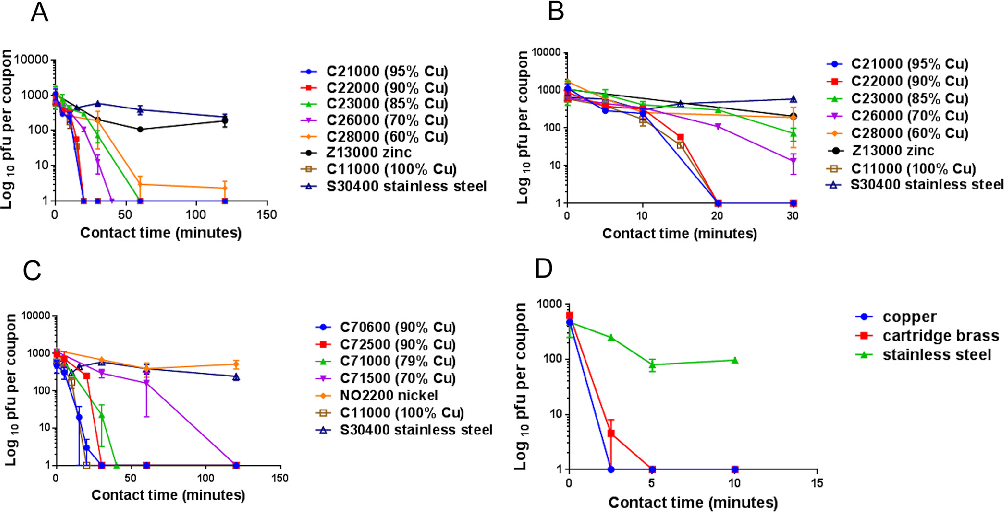
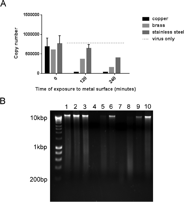
Viral titer decay curves on stainless steel and other metal surfaces demonstrate that coronavirus 229E can remain infectious for several days under ambient conditions.
Human Coronavirus 229E Remains Infectious on Common Touch Surface Materials.
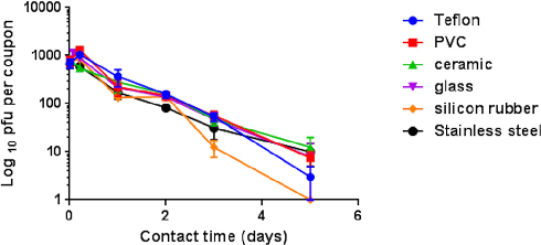
Comparison of coronavirus inactivation rates across polymer-based surfaces including PVC, silicone rubber, and Teflon shows material-dependent variation in viral survival.
Human Coronavirus 229E Remains Infectious on Common Touch Surface Materials.
Temperature and humidity effects on coronavirus 229E survival on surfaces are displayed, with higher temperatures generally associated with faster viral inactivation.
Human Coronavirus 229E Remains Infectious on Common Touch Surface Materials.
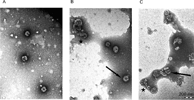
Scanning electron microscopy or surface characterization images of the tested materials are presented alongside their corresponding viral persistence data.
Human Coronavirus 229E Remains Infectious on Common Touch Surface Materials.
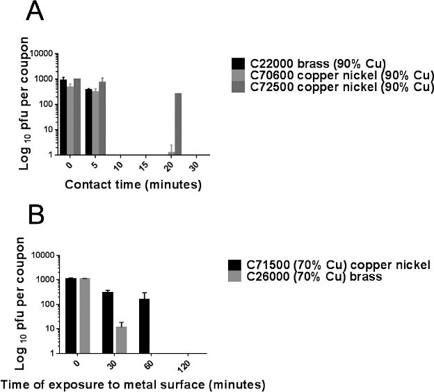
Recovery efficiency controls for the surface sampling methodology are shown, validating the quantitative viral titer measurements across different material types.
Human Coronavirus 229E Remains Infectious on Common Touch Surface Materials.
Summary comparison of coronavirus 229E persistence across all tested surface materials, ranked by duration of detectable infectivity.
Human Coronavirus 229E Remains Infectious on Common Touch Surface Materials.
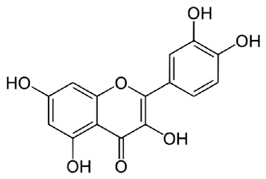
Chemical structure of quercetin (3,3',4',5,7-pentahydroxyflavone) is displayed, showing the hydroxyl groups on the flavonoid backbone that contribute to its antioxidant and anti-allergic properties.
Quercetin and Its Anti-Allergic Immune Response.
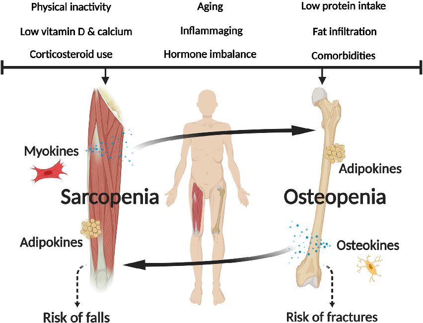
Pathophysiological mechanisms shared between osteoporosis and sarcopenia are diagrammed, including hormonal, nutritional, and mechanical loading factors.
Osteosarcopenia: epidemiology, diagnosis, and treatment-facts and numbers.
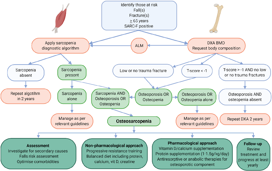
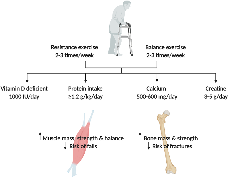
Treatment algorithms for osteosarcopenia incorporating exercise, nutritional supplementation, and pharmacological interventions are outlined.
Osteosarcopenia: epidemiology, diagnosis, and treatment-facts and numbers.
Outcome data from intervention studies targeting both bone and muscle health in osteosarcopenic patients are compared across different therapeutic approaches.
Osteosarcopenia: epidemiology, diagnosis, and treatment-facts and numbers.
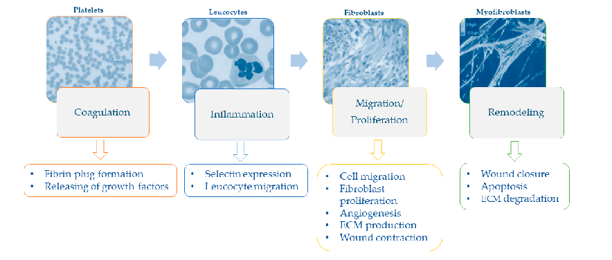
Sequential phases of the wound healing process - hemostasis, inflammation, proliferation, and remodeling - and their specific cellular events are illustrated.
Nutrition and Wound Healing: An Overview Focusing on the Beneficial Effects of …
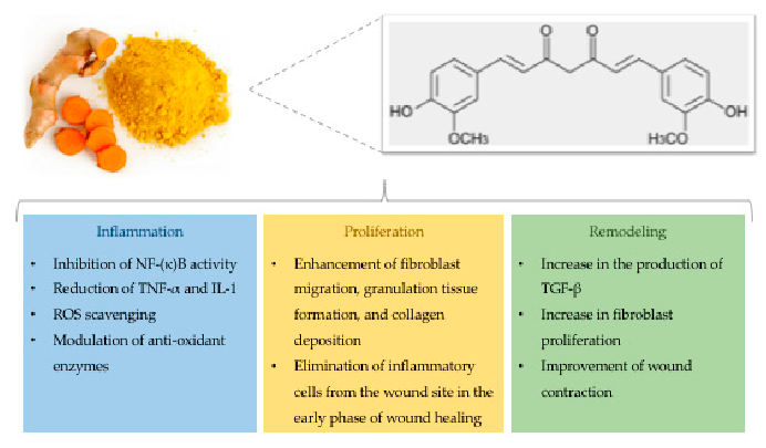
Chemical structure of curcumin alongside its documented effects on wound healing, including anti-inflammatory, antioxidant, and antimicrobial activities, are presented.
Nutrition and Wound Healing: An Overview Focusing on the Beneficial Effects of …
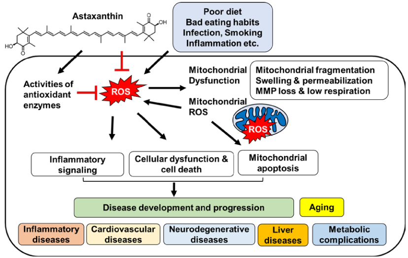
Proposed comprehensive mechanism by which astaxanthin inhibits oxidative stress-induced mitochondrial dysfunction, preventing downstream apoptotic and inflammatory signaling cascades.
Inhibitory Effect of Astaxanthin on Oxidative Stress-Induced Mitochondrial Dysfunction-A Mini-Review.
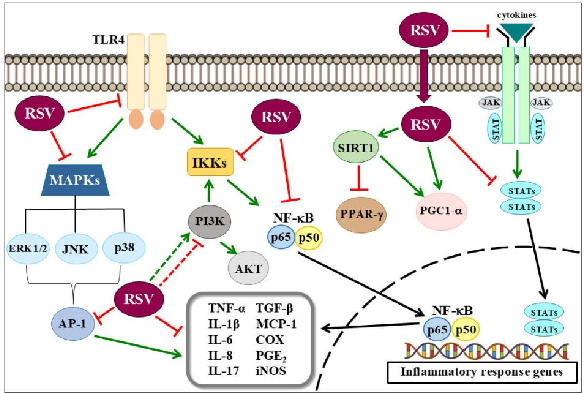
Molecular bases of resveratrol's anti-inflammatory effects are comprehensively illustrated, showing how inflammation activates multiple signaling pathways that are concurrently modulated by resveratrol through NF-kB, MAPK, and SIRT1 mechanisms.
Anti-Inflammatory Effects of Resveratrol: Mechanistic Insights.
Multiple convergent pathways leading to mitochondrial dysfunction in neuroimmune and neuropsychiatric disorders are mapped, including oxidative stress, calcium dysregulation, and inflammatory cytokine signaling.
The many roads to mitochondrial dysfunction in neuroimmune and neuropsychiatric disorders.
Gut microbiome composition alterations in RA patients and how dietary prebiotics and probiotics may influence disease progression.
Dietary Habits and Nutrition in Rheumatoid Arthritis: Can Diet Influence Disease Development …
Vitamin D metabolism and its immunomodulatory roles relevant to rheumatoid arthritis pathogenesis are diagrammed.
Dietary Habits and Nutrition in Rheumatoid Arthritis: Can Diet Influence Disease Development …
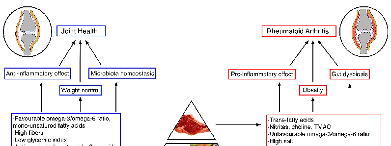
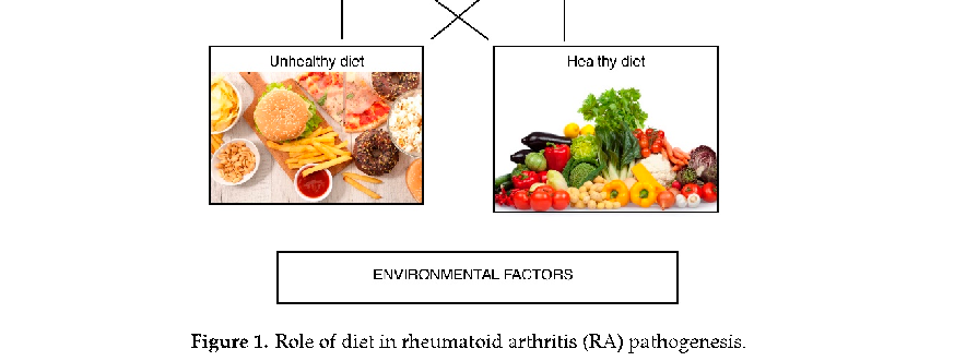
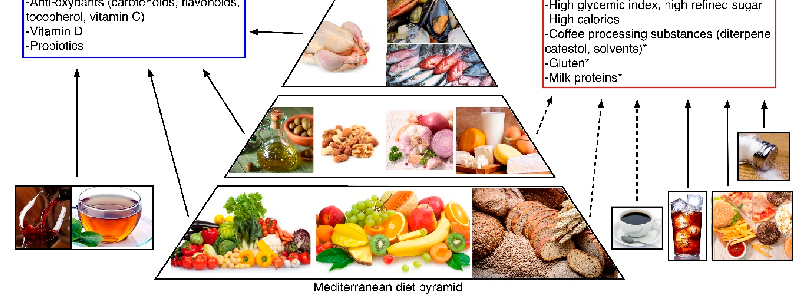
Nutrients and their food sources involved in RA development and progression are mapped, with asterisks indicating nutrients with less well-defined evidence for their role in the disease.
Dietary Habits and Nutrition in Rheumatoid Arthritis: Can Diet Influence Disease Development …
Polyphenol-rich foods and their documented anti-inflammatory mechanisms relevant to rheumatoid arthritis management are categorized.
Dietary Habits and Nutrition in Rheumatoid Arthritis: Can Diet Influence Disease Development …
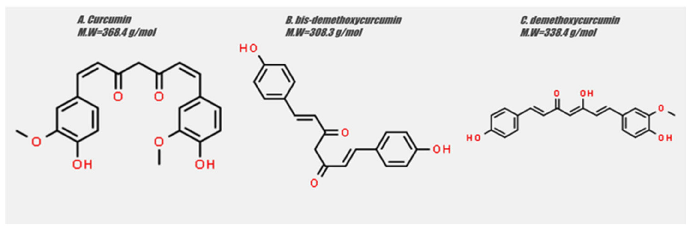
Chemical structures of curcumin, bis-demethoxycurcumin, and demethoxycurcumin - the three main curcuminoids - are displayed, highlighting the structural differences that influence their biological activity.
The Role of Curcumin in Cancer Treatment.
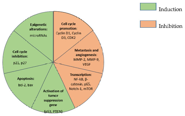
Summarized effects of curcumin on cancer cells are comprehensively mapped, showing modulation of CDK2, MMP-2, MMP-9, and multiple other molecular targets involved in tumor growth and metastasis.
The Role of Curcumin in Cancer Treatment.
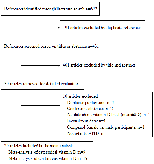
Meta-analysis results indicate that vitamin D deficiency is significantly more prevalent among patients with autoimmune thyroid disease compared to healthy controls. Lower serum 25-hydroxyvitamin D levels are associated with increased odds of both Hashimoto's thyroiditis and Graves' disease.
Meta-analysis of the association between vitamin D and autoimmune thyroid disease.
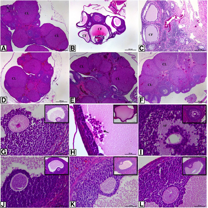
Histopathological analysis of ovarian sections across control and experimental groups shows characteristic PCOS features including multiple cystic follicles and thickened theca cell layers. Treatment groups demonstrate partial normalization of ovarian morphology.
Effect of resveratrol and metformin on ovarian reserve and ultrastructure in PCOS: …
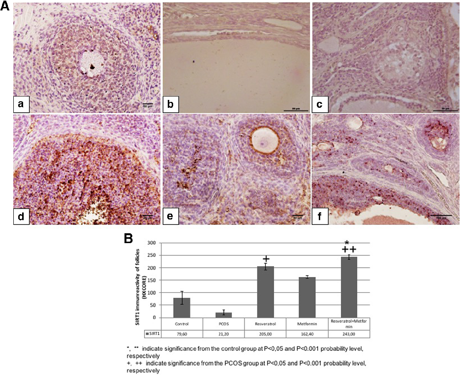
SIRT1 immunoreactivity in ovarian tissue varies across experimental groups, with reduced expression in PCOS rats and partial restoration following resveratrol and metformin treatment. SIRT1 is implicated in cellular stress responses and metabolic regulation.
Effect of resveratrol and metformin on ovarian reserve and ultrastructure in PCOS: …
Página 1 de 14