Deskripsi
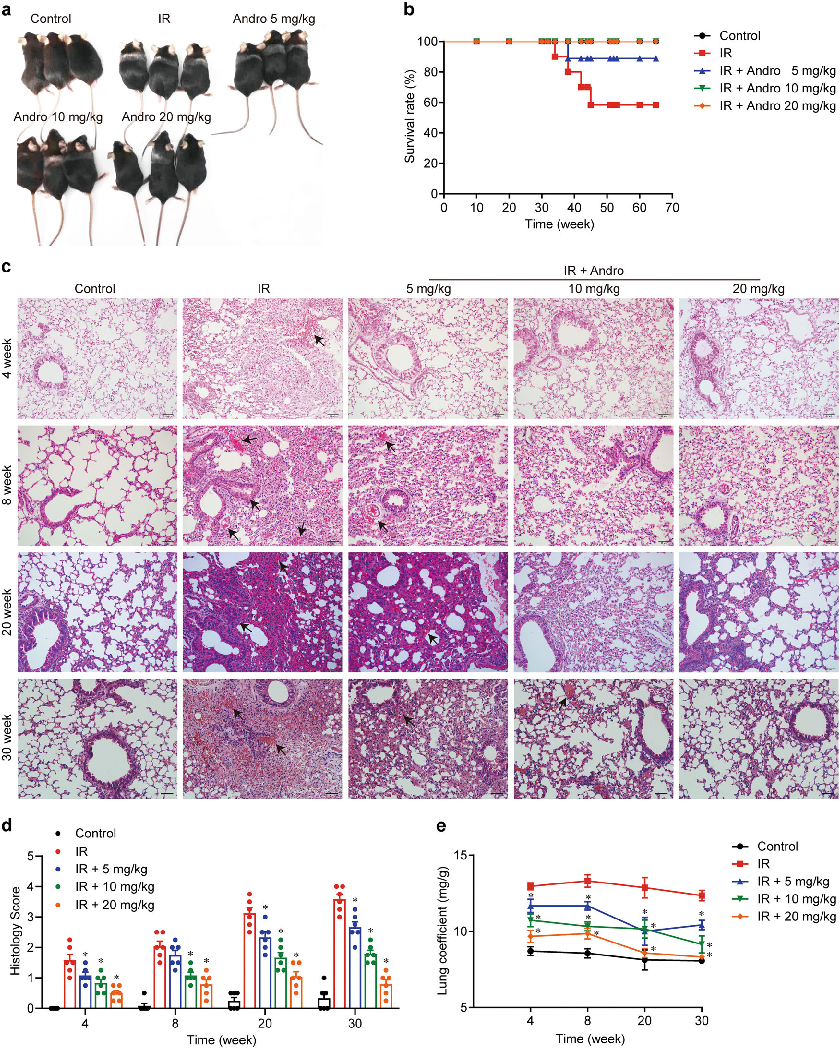
Mice exposed to 18 Gy irradiation and treated with varying doses of andrographolide for 4 weeks showed dose-dependent protection from radiation-induced lung injury. Representative images and quantitative data indicate that andrographolide significantly attenuated lung tissue damage.
More Figures from This Paper
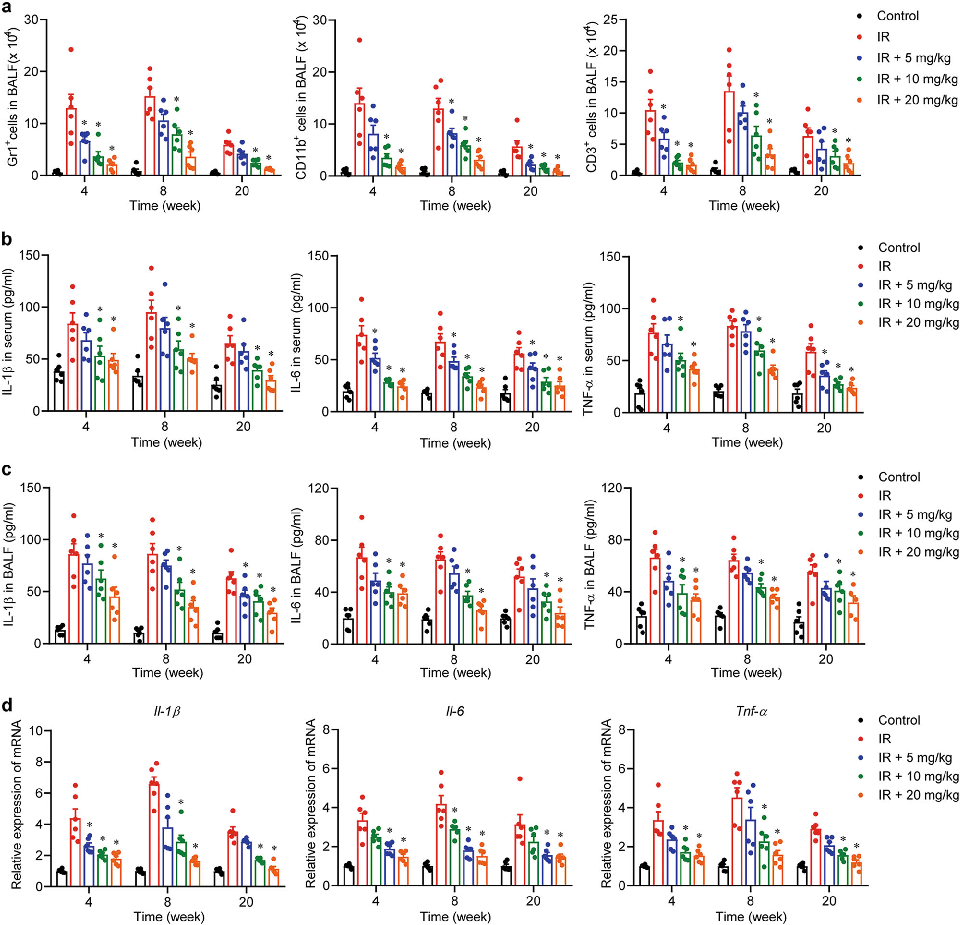
Figure 3
Histological analysis of lung tissue from irradiated mice reveals the extent of inflammatory cell infiltration and fibrosis. Andrographolide treatment appears to reduce these pathological changes in a dose-dependent manner.
micrograph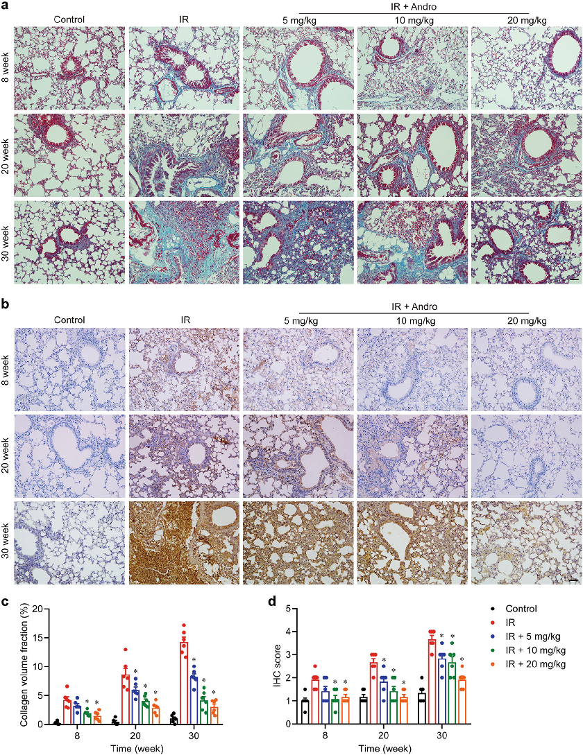
Figure 4
Inflammatory cytokine levels in lung tissue were measured following radiation exposure and andrographolide treatment. The data suggest that andrographolide suppresses pro-inflammatory mediator release in irradiated lung tissue.
chart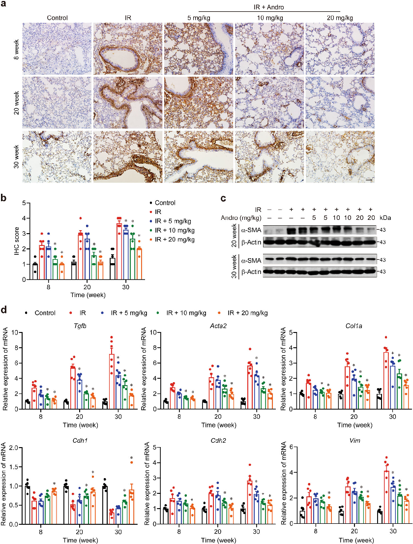
Figure 5
AIM2 inflammasome activation and caspase-1-mediated pyroptosis play key roles in radiation-induced lung inflammation. This figure presents protein expression data showing andrographolide's inhibitory effects on the AIM2 inflammasome pathway.
chart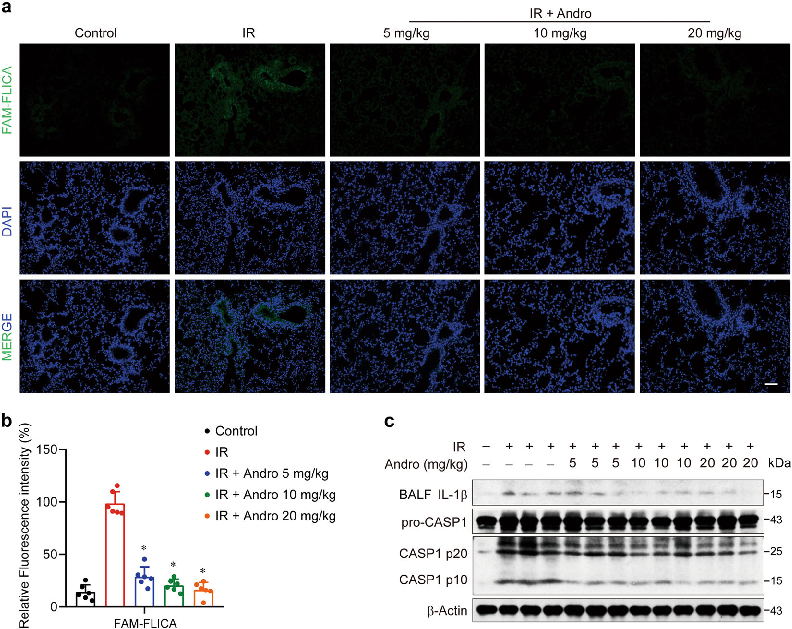
Figure 6
Gasdermin D cleavage is a downstream event in pyroptotic cell death triggered by radiation. Western blot analysis demonstrates that andrographolide reduces Gasdermin D processing in macrophages exposed to radiation.
chart
Figure 7
Immunofluorescence or protein analysis reveals that andrographolide prevents AIM2 from binding cytoplasmic DNA and forming active inflammasome complexes. These findings indicate a specific molecular target for andrographolide's protective mechanism.
chart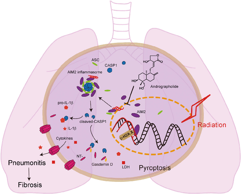
Figure 8
Andrographolide ameliorates radiation-induced lung injury by inhibiting Caspase-1-mediated Gasdermin D-dependent pyroptosis in macrophages. This schematic illustrates how the compound prevents AIM2 from translocating into the nucleus, thereby blocking inflammasome assembly and downstream inflammatory cascades.
diagramFigure 2
PhotographSource Paper
Inhibition of AIM2 inflammasome-mediated pyroptosis by Andrographolide contributes to amelioration of radiation-induced lung inflammation and fibrosis.Cite This Figure
 > Source: Jian Gao et al. "Inhibition of AIM2 inflammasome-mediated pyroptosis by Andrographolide contribut." *Cell death & disease*, 2019. PMID: [31862870](https://pubmed.ncbi.nlm.nih.gov/31862870/)
<figure>
  <img src="https://pdfs.citedhealth.com/figures/31862870/150.png" alt="Mice exposed to 18 Gy irradiation and treated with varying doses of andrographolide for 4 weeks showed dose-dependent protection from radiation-induced lung injury. Representative images and quantitative data indicate that andrographolide significantly attenuated lung tissue damage." />
  <figcaption>Figure 2. Mice exposed to 18 Gy irradiation and treated with varying doses of andrographolide for 4 weeks showed dose-dependent protection from radiation-induced lung injury. Representative images and quantitative data indicate that andrographolide significantly attenuated lung tissue damage.<br>  Source: Jian Gao et al. "Inhibition of AIM2 inflammasome-mediated pyroptosis by Andrographolide contribut." <em>Cell death & disease</em>, 2019. PMID: <a href="https://pubmed.ncbi.nlm.nih.gov/31862870/">31862870</a></figcaption>
</figure>