Deskripsi
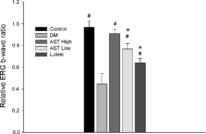
Electroretinography (ERG) recordings evaluating retinal function in control and STZ-induced diabetic rats treated with normal saline, 0.6 mg/kg AST, 3 mg/kg AST, or 0.5 mg/kg lutein for 8 weeks. ERG wave amplitudes indicate the functional impact of each treatment on diabetic retinal physiology.
More Figures from This Paper
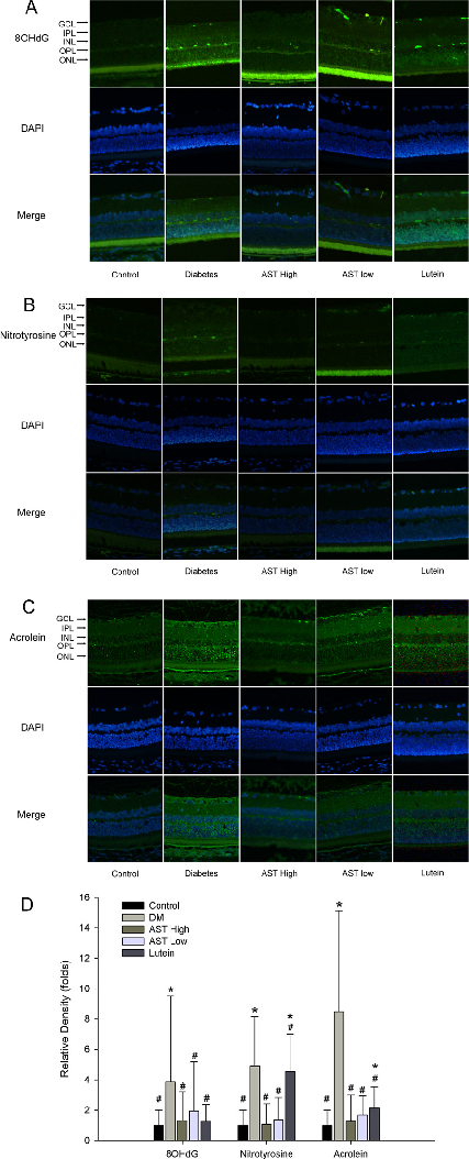
Figure 5
Inflammatory marker expression levels in retinal tissue or aqueous humor of diabetic rats, comparing astaxanthin-treated groups with untreated diabetic controls. Results indicate AST may modulate inflammatory mediator production.
chart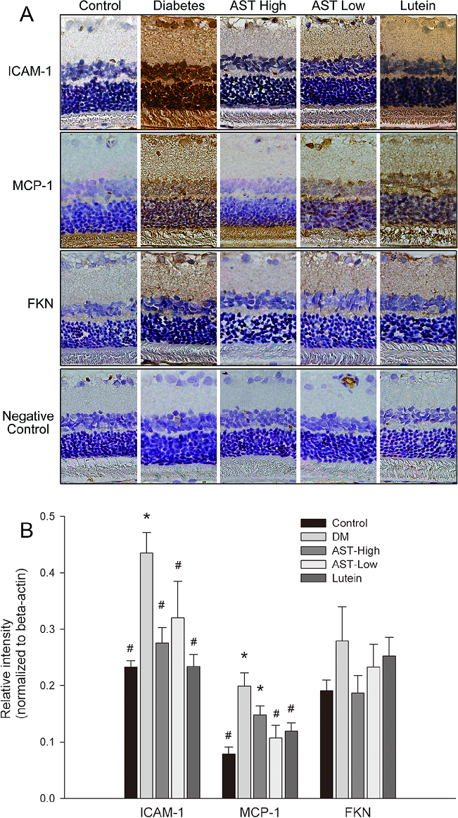
Figure 6
Western blot or protein expression analysis of retinal inflammatory and oxidative stress markers in the diabetic rat model, demonstrating the molecular effects of astaxanthin treatment on key signaling proteins.
chart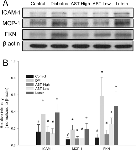
Figure 7
Gene or protein expression data for retinal vascular endothelial growth factor (VEGF) or related angiogenic markers in AST-treated versus untreated diabetic rats.
chart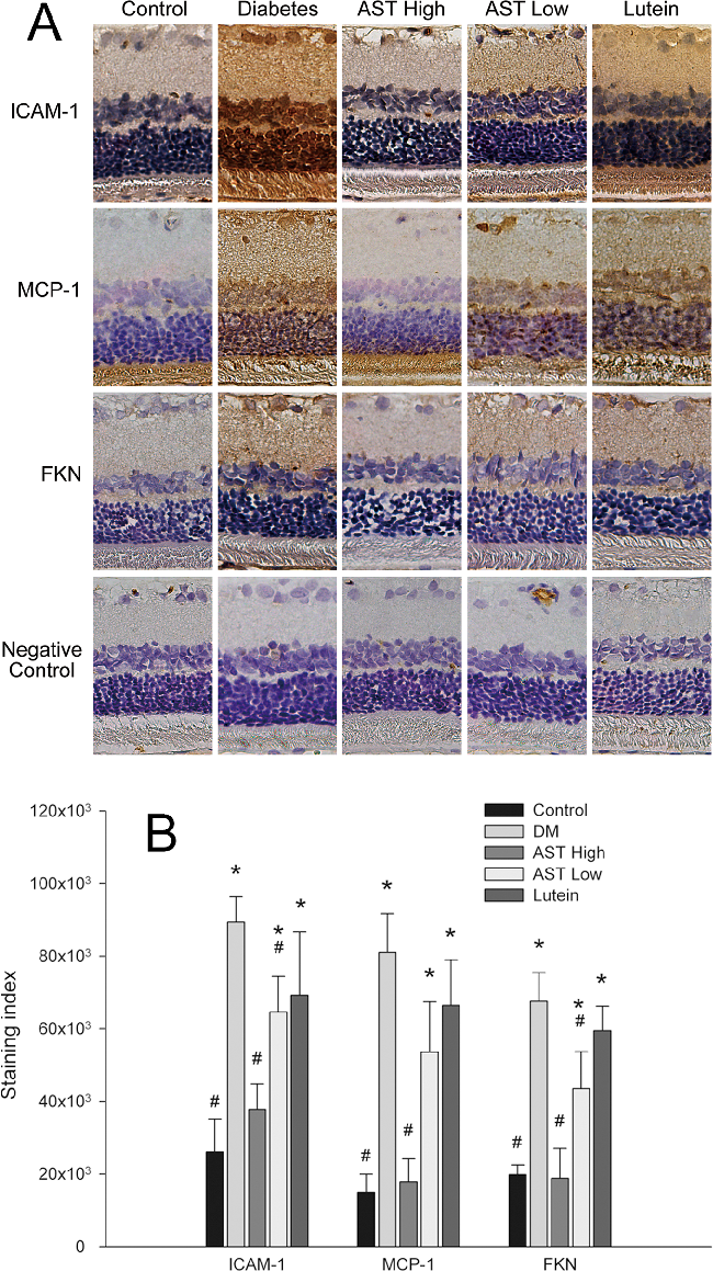
Figure 8
Histological or immunohistochemical analysis of retinal tissue sections from diabetic rats, comparing structural changes across treatment groups to assess astaxanthin's protective effects on retinal architecture.
micrograph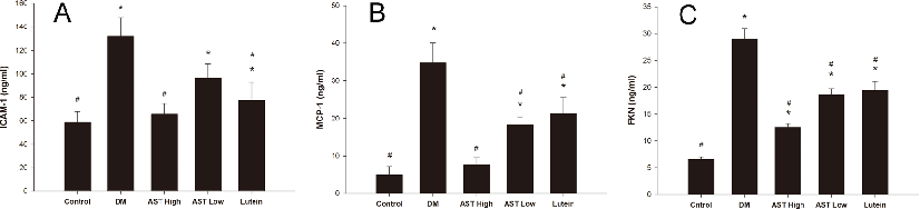
Figure 9
Quantification of ICAM-1, MCP-1, and fractalkine (FKN) protein levels in aqueous humor of diabetic rats treated with astaxanthin or lutein. Reduced expression of these inflammatory mediators suggests AST may attenuate vascular inflammation in diabetic eyes.
chart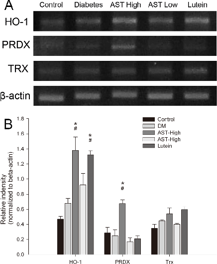
Figure 10
Supplementary analysis of inflammatory or oxidative markers in ocular tissues of STZ-induced diabetic rats, providing additional evidence for astaxanthin's protective mechanism against diabetic retinal damage.
chartFigure 4
ChartSource Paper
Astaxanthin Inhibits Expression of Retinal Oxidative Stress and Inflammatory Mediators in Streptozotocin-Induced Diabetic Rats.Cite This Figure
 > Source: Po-Ting Yeh et al. "Astaxanthin Inhibits Expression of Retinal Oxidative Stress and Inflammatory Med." *PloS one*, 2016. PMID: [26765843](https://pubmed.ncbi.nlm.nih.gov/26765843/)
<figure>
  <img src="https://pdfs.citedhealth.com/figures/26765843/235.png" alt="Electroretinography (ERG) recordings evaluating retinal function in control and STZ-induced diabetic rats treated with normal saline, 0.6 mg/kg AST, 3 mg/kg AST, or 0.5 mg/kg lutein for 8 weeks. ERG wave amplitudes indicate the functional impact of each treatment on diabetic retinal physiology." />
  <figcaption>Figure 4. Electroretinography (ERG) recordings evaluating retinal function in control and STZ-induced diabetic rats treated with normal saline, 0.6 mg/kg AST, 3 mg/kg AST, or 0.5 mg/kg lutein for 8 weeks. ERG wave amplitudes indicate the functional impact of each treatment on diabetic retinal physiology.<br>  Source: Po-Ting Yeh et al. "Astaxanthin Inhibits Expression of Retinal Oxidative Stress and Inflammatory Med." <em>PloS one</em>, 2016. PMID: <a href="https://pubmed.ncbi.nlm.nih.gov/26765843/">26765843</a></figcaption>
</figure>