Resveratrol الأشكال
6 أشكال من أبحاث محكّمة
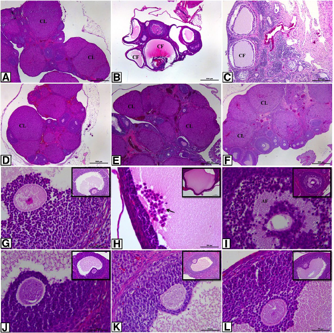
Histopathological analysis of ovarian sections across control and experimental groups shows characteristic PCOS features including multiple cystic follicles and thickened theca cell layers. Treatment groups demonstrate partial normalization of ovarian morphology.
Effect of resveratrol and metformin on ovarian reserve and ultrastructure in PCOS: …
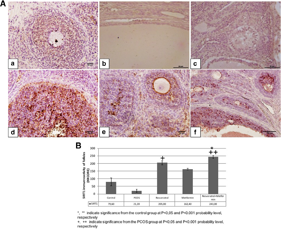
SIRT1 immunoreactivity in ovarian tissue varies across experimental groups, with reduced expression in PCOS rats and partial restoration following resveratrol and metformin treatment. SIRT1 is implicated in cellular stress responses and metabolic regulation.
Effect of resveratrol and metformin on ovarian reserve and ultrastructure in PCOS: …
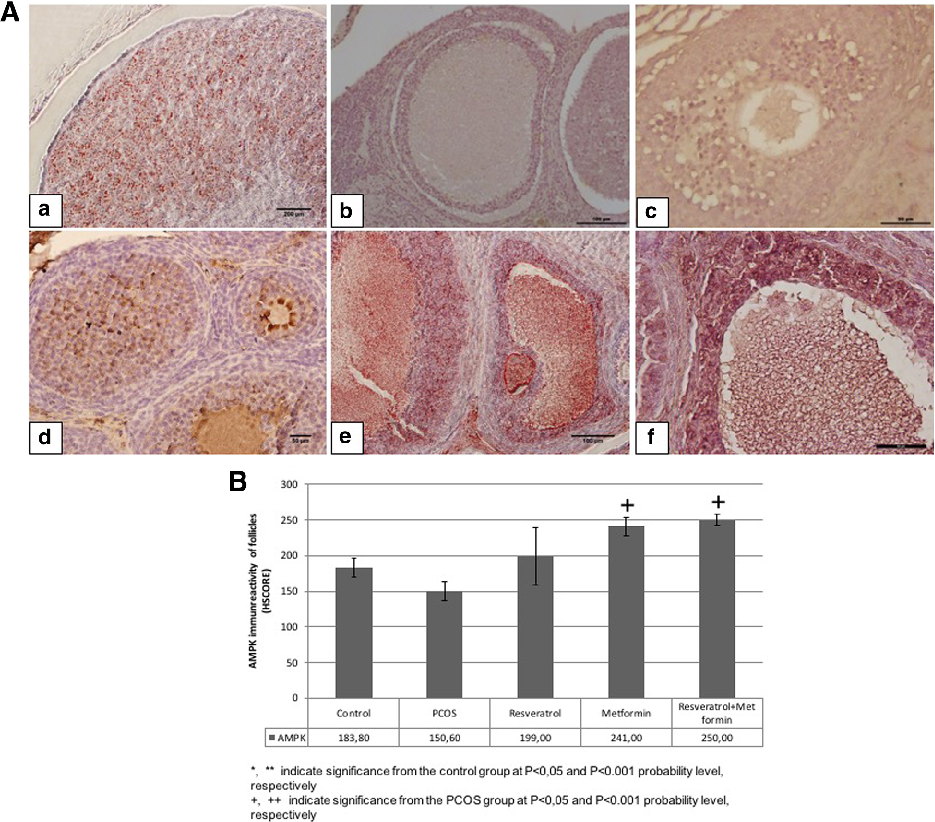
AMPK immunoreactivity analysis reveals decreased phosphorylated AMPK in PCOS ovarian tissue compared to controls. Both metformin and resveratrol treatments partially restore AMPK activation, consistent with their known metabolic signaling effects.
Effect of resveratrol and metformin on ovarian reserve and ultrastructure in PCOS: …
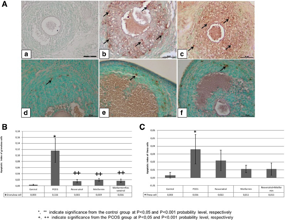
TUNEL analysis of granulosa and theca cells quantifies apoptotic rates across experimental groups. PCOS induction increases apoptosis in granulosa cells, while resveratrol and metformin treatments reduce apoptotic indices toward control levels.
Effect of resveratrol and metformin on ovarian reserve and ultrastructure in PCOS: …
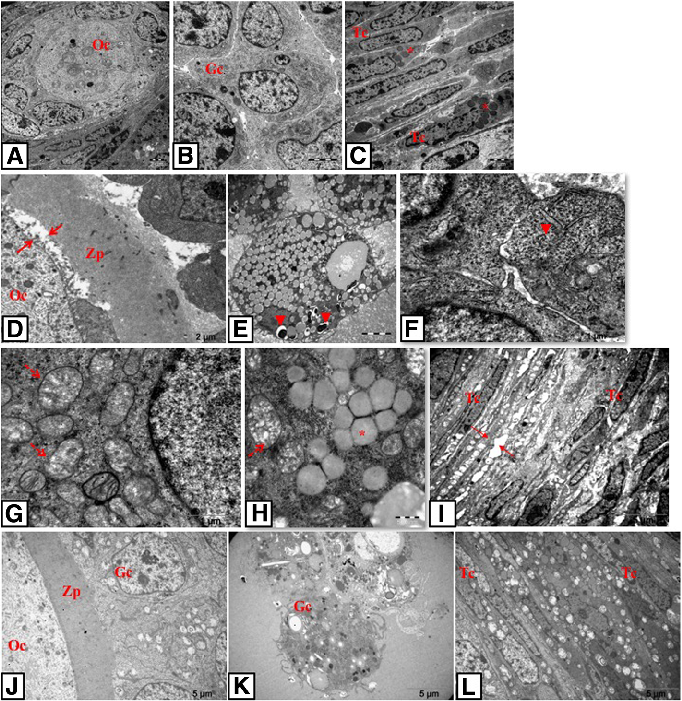
Transmission electron microscopy photomicrographs reveal ultrastructural details of oocytes, granulosa cells, and theca cells in control and PCOS groups. PCOS follicles exhibit mitochondrial swelling, dilated endoplasmic reticulum, and other signs of cellular stress.
Effect of resveratrol and metformin on ovarian reserve and ultrastructure in PCOS: …
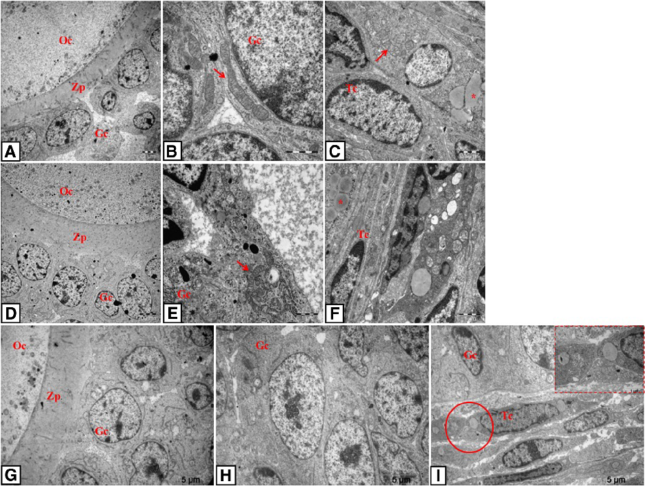
TEM photomicrographs of resveratrol-, metformin-, and combination-treated rat ovarian tissue show improved ultrastructural features compared to untreated PCOS. Mitochondrial morphology and endoplasmic reticulum integrity are partially restored.
Effect of resveratrol and metformin on ovarian reserve and ultrastructure in PCOS: …