Astaxanthin Gambar
28 gambar dari penelitian yang ditinjau oleh rekan sejawat
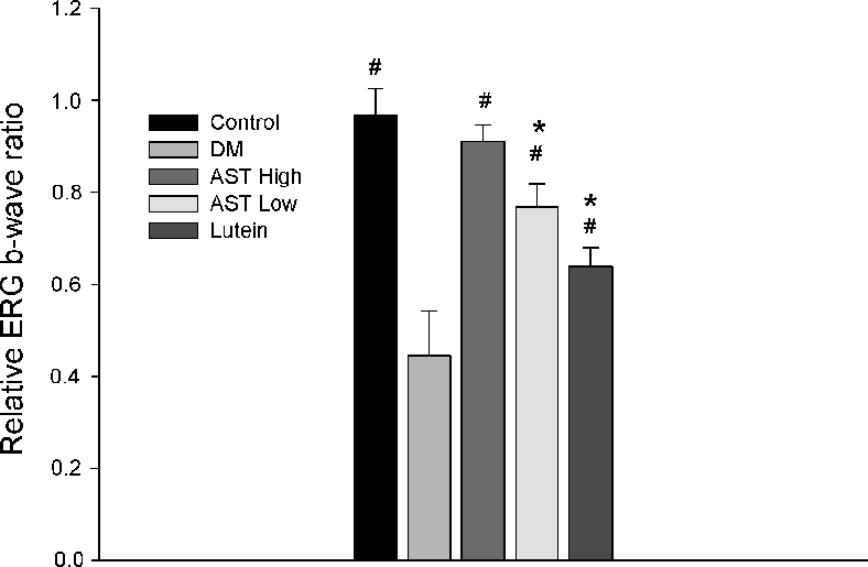
Electroretinography (ERG) recordings evaluating retinal function in control and STZ-induced diabetic rats treated with normal saline, 0.6 mg/kg AST, 3 mg/kg AST, or 0.5 mg/kg lutein for 8 weeks. ERG wave amplitudes indicate the functional impact of each treatment on diabetic retinal physiology.
Astaxanthin Inhibits Expression of Retinal Oxidative Stress and Inflammatory Mediators in Streptozotocin-Induced …
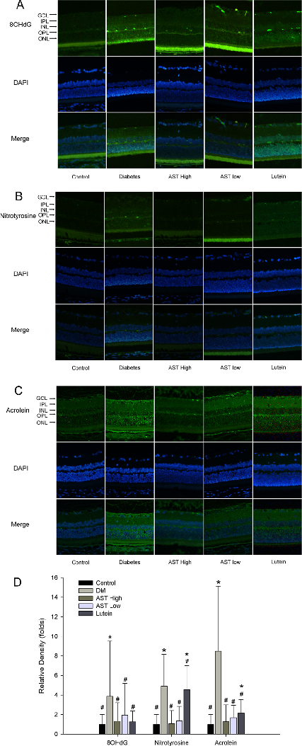
Inflammatory marker expression levels in retinal tissue or aqueous humor of diabetic rats, comparing astaxanthin-treated groups with untreated diabetic controls. Results indicate AST may modulate inflammatory mediator production.
Astaxanthin Inhibits Expression of Retinal Oxidative Stress and Inflammatory Mediators in Streptozotocin-Induced …
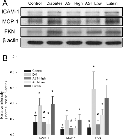
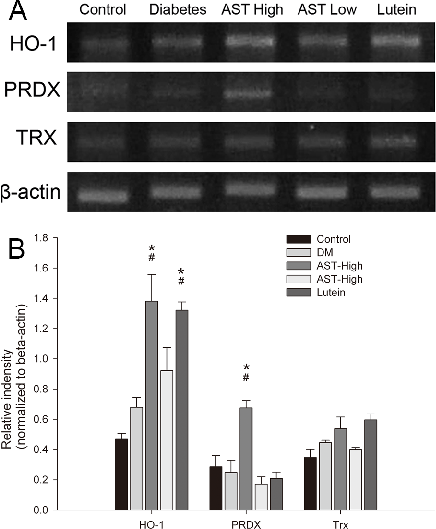
Western blot or protein expression analysis of retinal inflammatory and oxidative stress markers in the diabetic rat model, demonstrating the molecular effects of astaxanthin treatment on key signaling proteins.
Astaxanthin Inhibits Expression of Retinal Oxidative Stress and Inflammatory Mediators in Streptozotocin-Induced …
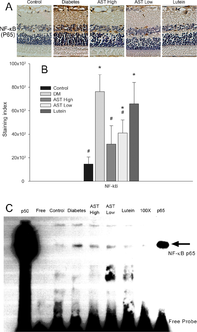
Gene or protein expression data for retinal vascular endothelial growth factor (VEGF) or related angiogenic markers in AST-treated versus untreated diabetic rats.
Astaxanthin Inhibits Expression of Retinal Oxidative Stress and Inflammatory Mediators in Streptozotocin-Induced …
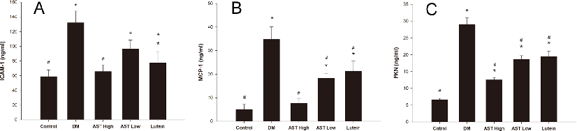
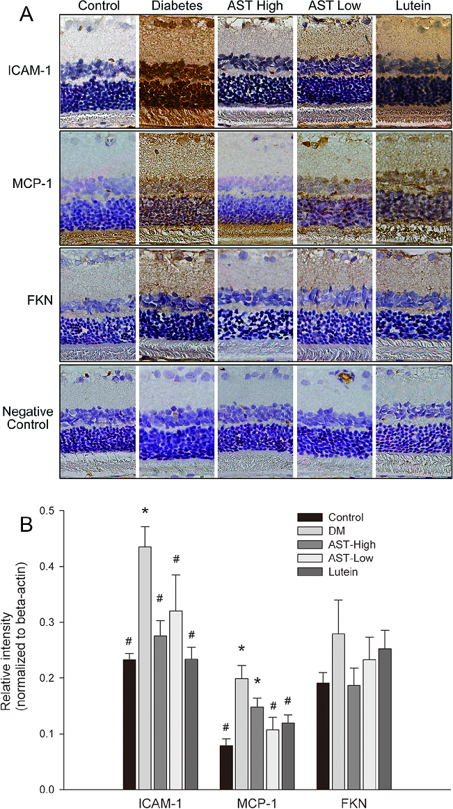
Quantification of ICAM-1, MCP-1, and fractalkine (FKN) protein levels in aqueous humor of diabetic rats treated with astaxanthin or lutein. Reduced expression of these inflammatory mediators suggests AST may attenuate vascular inflammation in diabetic eyes.
Astaxanthin Inhibits Expression of Retinal Oxidative Stress and Inflammatory Mediators in Streptozotocin-Induced …
Supplementary analysis of inflammatory or oxidative markers in ocular tissues of STZ-induced diabetic rats, providing additional evidence for astaxanthin's protective mechanism against diabetic retinal damage.
Astaxanthin Inhibits Expression of Retinal Oxidative Stress and Inflammatory Mediators in Streptozotocin-Induced …
Summary of retinal protection outcomes or dose-response data for astaxanthin in the diabetic rat model, consolidating evidence that AST inhibits expression of oxidative stress and inflammatory mediators in diabetic retinopathy.
Astaxanthin Inhibits Expression of Retinal Oxidative Stress and Inflammatory Mediators in Streptozotocin-Induced …
Experimental results examining simultaneous Inhibitory Effects of All, with data points illustrating key findings related to alzheimer´s disease is a global neurodegenerative health concern.
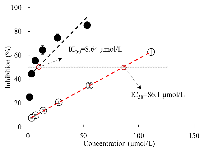
Simultaneous Inhibitory Effects of All-Trans Astaxanthin on Acetylcholinesterase and Oxidative Stress.
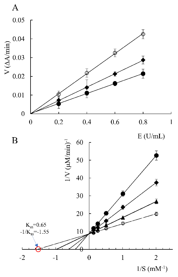
Reaction rates of acetylcholinesterase in the presence of all-trans astaxanthin with different concentrations (A), and Lineweaver–Burk reciprocal plots (B). ○: all-trans astaxanthin concentration 0 μmol/L; ▲: all-trans astaxanthin concentration 6.5 μmol/L; ◆: all-trans astaxanthin concentration 26 μmol/L; ●: all-trans astaxanthin concentration 5...
Simultaneous Inhibitory Effects of All-Trans Astaxanthin on Acetylcholinesterase and Oxidative Stress.
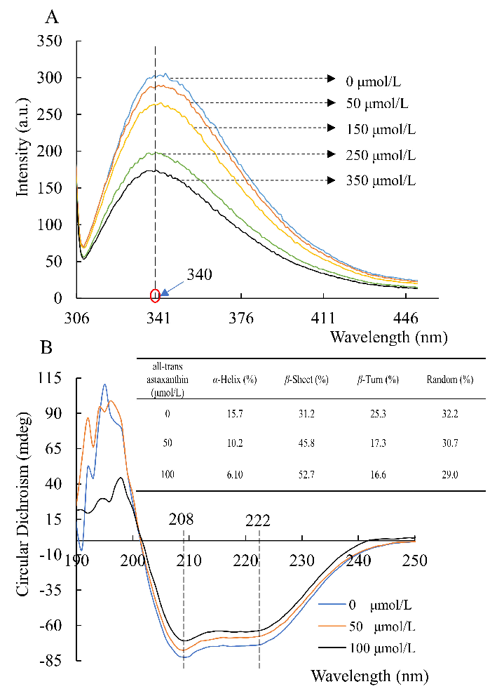
Fluorescence emission (A) and circular dichroism (B) spectra of acetylcholinesterase in the presence of all-trans astaxanthin with various concentrations.
Simultaneous Inhibitory Effects of All-Trans Astaxanthin on Acetylcholinesterase and Oxidative Stress.
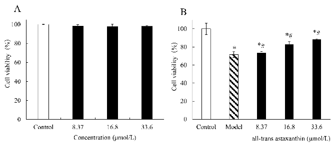
The effect of all-trans astaxanthin concentrations on cell viability in group (A) and in group (B) treated with Aβ25–35. : Control group; : astaxanthin-treatment group;
Simultaneous Inhibitory Effects of All-Trans Astaxanthin on Acetylcholinesterase and Oxidative Stress.
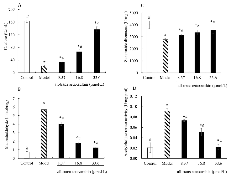
Effect of all-trans astaxanthin on intracellular antioxidant capacity and acetylcholinesterase activity. The levels of catalase (A), malondialdehyde (B), superoxide dismutase (C), and acetylcholinesterase activity (D) are shown.
Simultaneous Inhibitory Effects of All-Trans Astaxanthin on Acetylcholinesterase and Oxidative Stress.
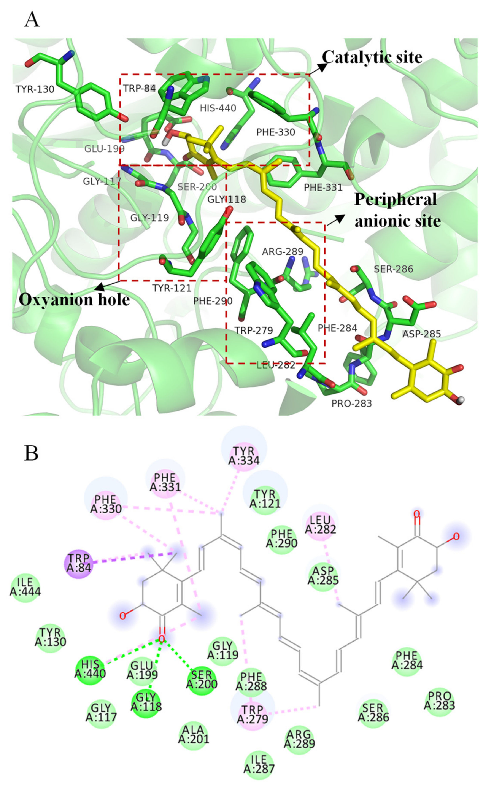
3D (A) and 2D (B) structural simulation of all-trans astaxanthin interacting with acetylcholinesterase.
Simultaneous Inhibitory Effects of All-Trans Astaxanthin on Acetylcholinesterase and Oxidative Stress.
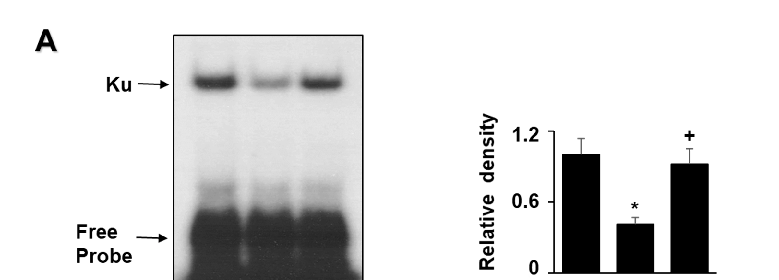
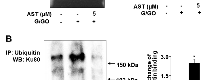
Experimental results examining astaxanthin Inhibits Oxidative Stress, with data points illustrating key findings related to oxidative stress induces DNA damage which can be repaired by DNA repair proteins, such as Ku70/80.
Astaxanthin Inhibits Oxidative Stress-Induced Ku Protein Degradation and Apoptosis in Gastric Epithelial …
Statistical analysis from research investigating astaxanthin Inhibits Oxidative Stress, comparing treatment groups and control conditions.
Astaxanthin Inhibits Oxidative Stress-Induced Ku Protein Degradation and Apoptosis in Gastric Epithelial …
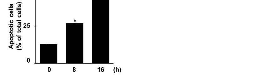
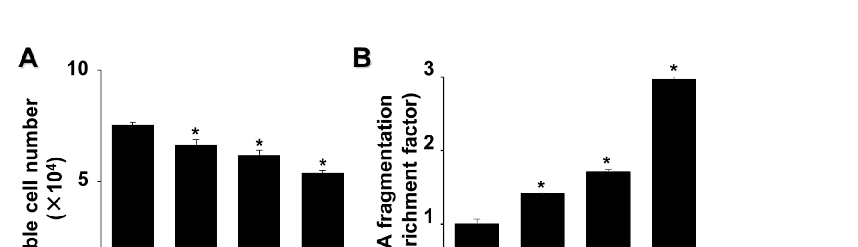
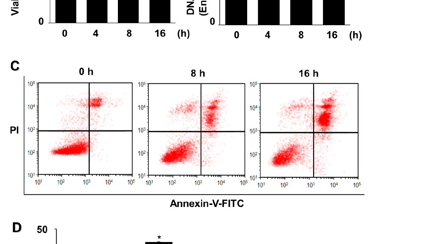
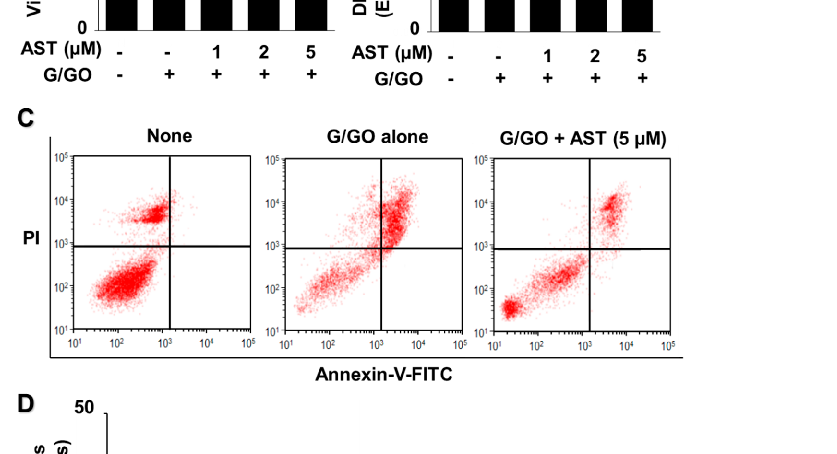
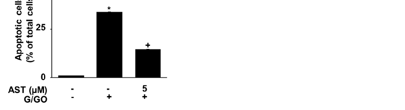
Glucose/glucose oxidase (G/GO) treatment induced cell death, DNA fragmentation, and apoptosis in AGS cells. The cells were stimulated with G/GO for the indicated periods.
Astaxanthin Inhibits Oxidative Stress-Induced Ku Protein Degradation and Apoptosis in Gastric Epithelial …
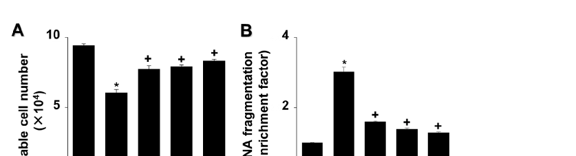
cells in G/GO-treated cells (Figure 2C,D). These data show that astaxanthin inhibits G/GOinduced cell death, DNA fragmentation, and apoptosis in AGS cells in a dose-dependent manner.
Astaxanthin Inhibits Oxidative Stress-Induced Ku Protein Degradation and Apoptosis in Gastric Epithelial …
Quantitative data from a study on astaxanthin Inhibits Oxidative Stress, presenting measured outcomes relevant to the investigation of oxidative stress induces DNA damage which can be repaired by DNA repair proteins, such as Ku70/80.
Astaxanthin Inhibits Oxidative Stress-Induced Ku Protein Degradation and Apoptosis in Gastric Epithelial …
Experimental results examining astaxanthin Inhibits Oxidative Stress, with data points illustrating key findings related to oxidative stress induces DNA damage which can be repaired by DNA repair proteins, such as Ku70/80.
Astaxanthin Inhibits Oxidative Stress-Induced Ku Protein Degradation and Apoptosis in Gastric Epithelial …
Statistical analysis from research investigating astaxanthin Inhibits Oxidative Stress, comparing treatment groups and control conditions.
Astaxanthin Inhibits Oxidative Stress-Induced Ku Protein Degradation and Apoptosis in Gastric Epithelial …
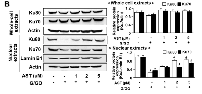
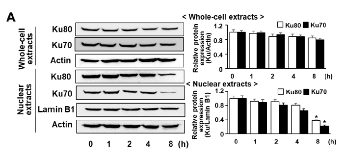
Astaxanthin inhibits glucose/glucose oxidase (G/GO)−induced decrease in Ku70/80 levels in whole-cell extracts and nuclear extracts in AGS cells. The cells were (A) treated with G/GO for the indicated time periods or (B) pre−treated with the indicated concentrations of astaxanthin for 3 h, followed by stimulation with G/GO for 8 h.
Astaxanthin Inhibits Oxidative Stress-Induced Ku Protein Degradation and Apoptosis in Gastric Epithelial …
Graphical representation of outcomes in a study of astaxanthin Inhibits Oxidative Stress, highlighting trends observed across experimental conditions.
Astaxanthin Inhibits Oxidative Stress-Induced Ku Protein Degradation and Apoptosis in Gastric Epithelial …
Quantitative data from a study on astaxanthin Inhibits Oxidative Stress, presenting measured outcomes relevant to the investigation of oxidative stress induces DNA damage which can be repaired by DNA repair proteins, such as Ku70/80.
Astaxanthin Inhibits Oxidative Stress-Induced Ku Protein Degradation and Apoptosis in Gastric Epithelial …
Experimental results examining astaxanthin Inhibits Oxidative Stress, with data points illustrating key findings related to oxidative stress induces DNA damage which can be repaired by DNA repair proteins, such as Ku70/80.
Astaxanthin Inhibits Oxidative Stress-Induced Ku Protein Degradation and Apoptosis in Gastric Epithelial …
Halaman 1 dari 2