Processo di ricerca
13 figure da ricerca revisionata da esperti
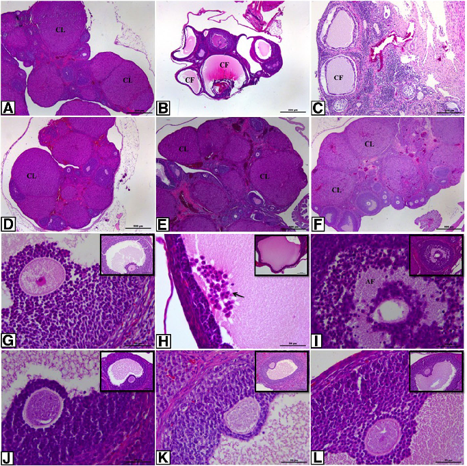
Histopathological analysis of ovarian sections across control and experimental groups shows characteristic PCOS features including multiple cystic follicles and thickened theca cell layers. Treatment groups demonstrate partial normalization of ovarian morphology.
Effect of resveratrol and metformin on ovarian reserve and ultrastructure in PCOS: …
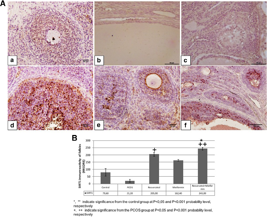
SIRT1 immunoreactivity in ovarian tissue varies across experimental groups, with reduced expression in PCOS rats and partial restoration following resveratrol and metformin treatment. SIRT1 is implicated in cellular stress responses and metabolic regulation.
Effect of resveratrol and metformin on ovarian reserve and ultrastructure in PCOS: …
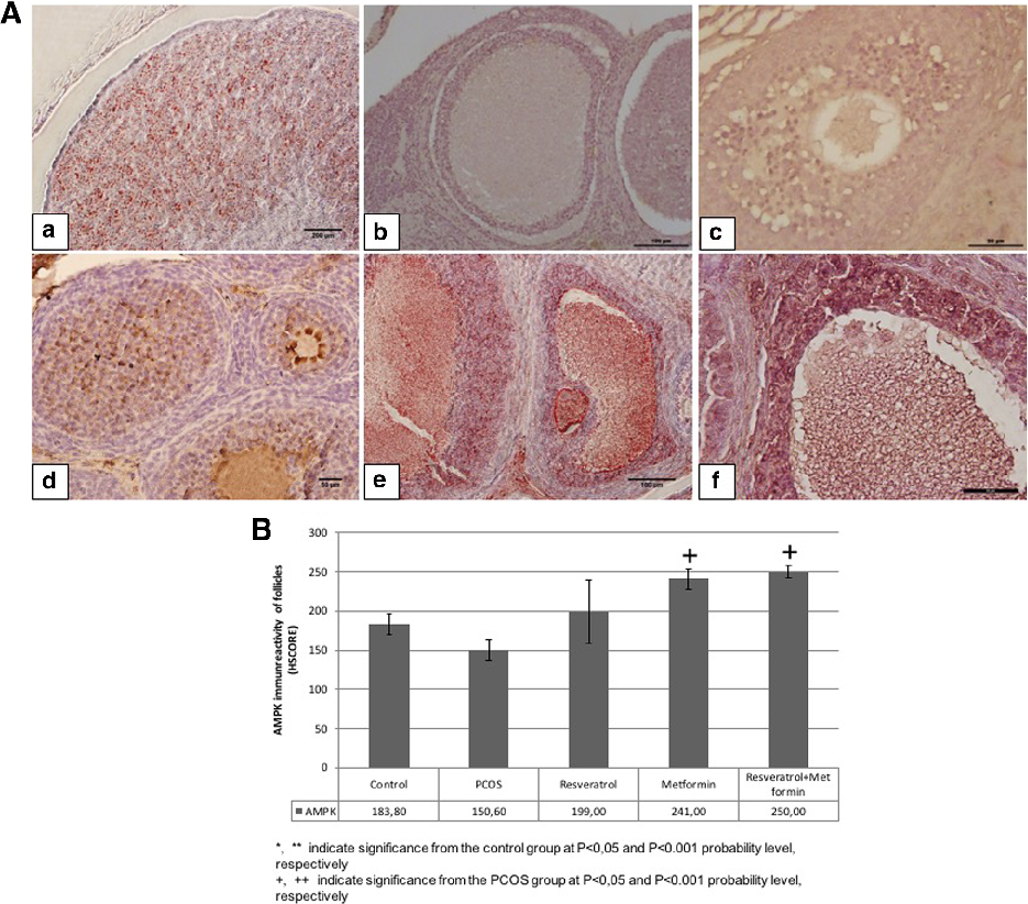
AMPK immunoreactivity analysis reveals decreased phosphorylated AMPK in PCOS ovarian tissue compared to controls. Both metformin and resveratrol treatments partially restore AMPK activation, consistent with their known metabolic signaling effects.
Effect of resveratrol and metformin on ovarian reserve and ultrastructure in PCOS: …
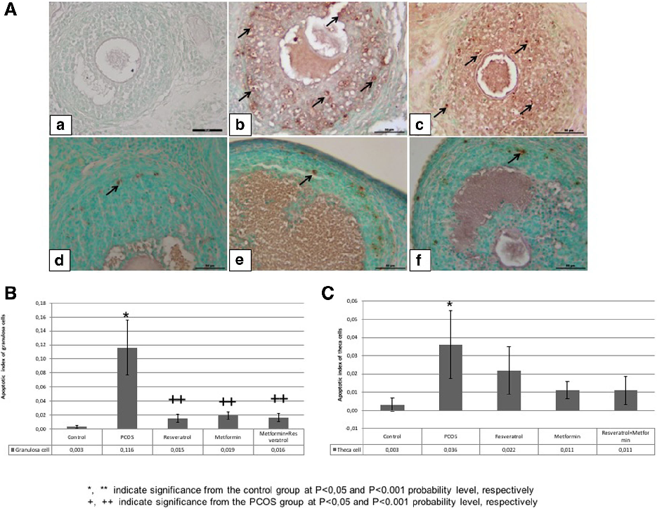
TUNEL analysis of granulosa and theca cells quantifies apoptotic rates across experimental groups. PCOS induction increases apoptosis in granulosa cells, while resveratrol and metformin treatments reduce apoptotic indices toward control levels.
Effect of resveratrol and metformin on ovarian reserve and ultrastructure in PCOS: …
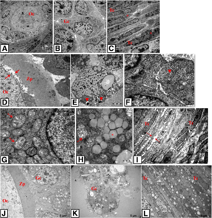
Transmission electron microscopy photomicrographs reveal ultrastructural details of oocytes, granulosa cells, and theca cells in control and PCOS groups. PCOS follicles exhibit mitochondrial swelling, dilated endoplasmic reticulum, and other signs of cellular stress.
Effect of resveratrol and metformin on ovarian reserve and ultrastructure in PCOS: …
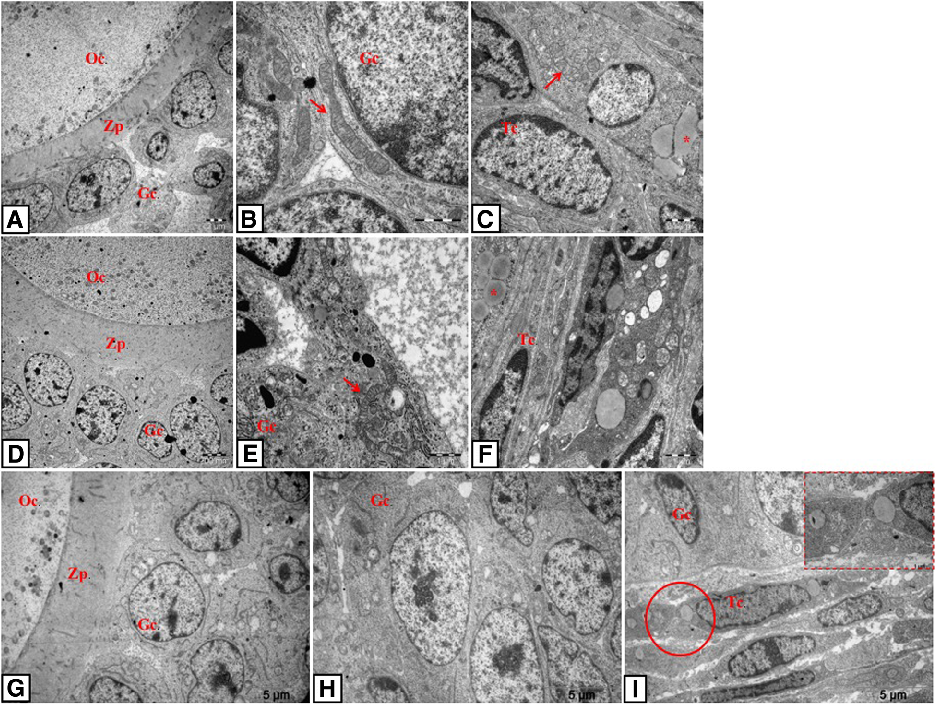
TEM photomicrographs of resveratrol-, metformin-, and combination-treated rat ovarian tissue show improved ultrastructural features compared to untreated PCOS. Mitochondrial morphology and endoplasmic reticulum integrity are partially restored.
Effect of resveratrol and metformin on ovarian reserve and ultrastructure in PCOS: …
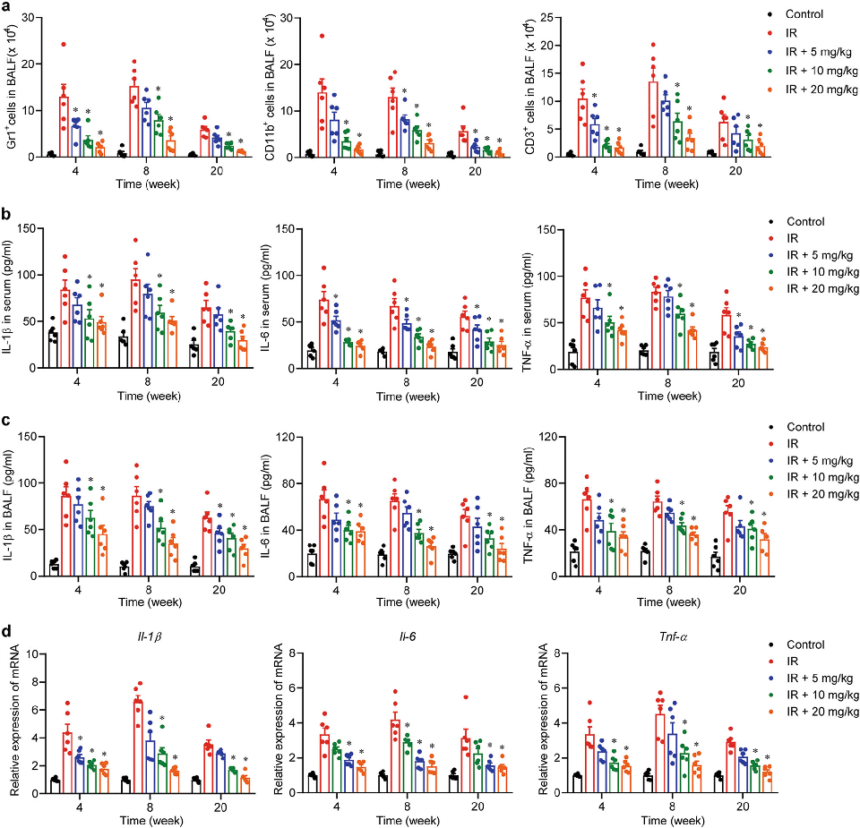
Histological analysis of lung tissue from irradiated mice reveals the extent of inflammatory cell infiltration and fibrosis. Andrographolide treatment appears to reduce these pathological changes in a dose-dependent manner.
Inhibition of AIM2 inflammasome-mediated pyroptosis by Andrographolide contributes to amelioration of radiation-induced …
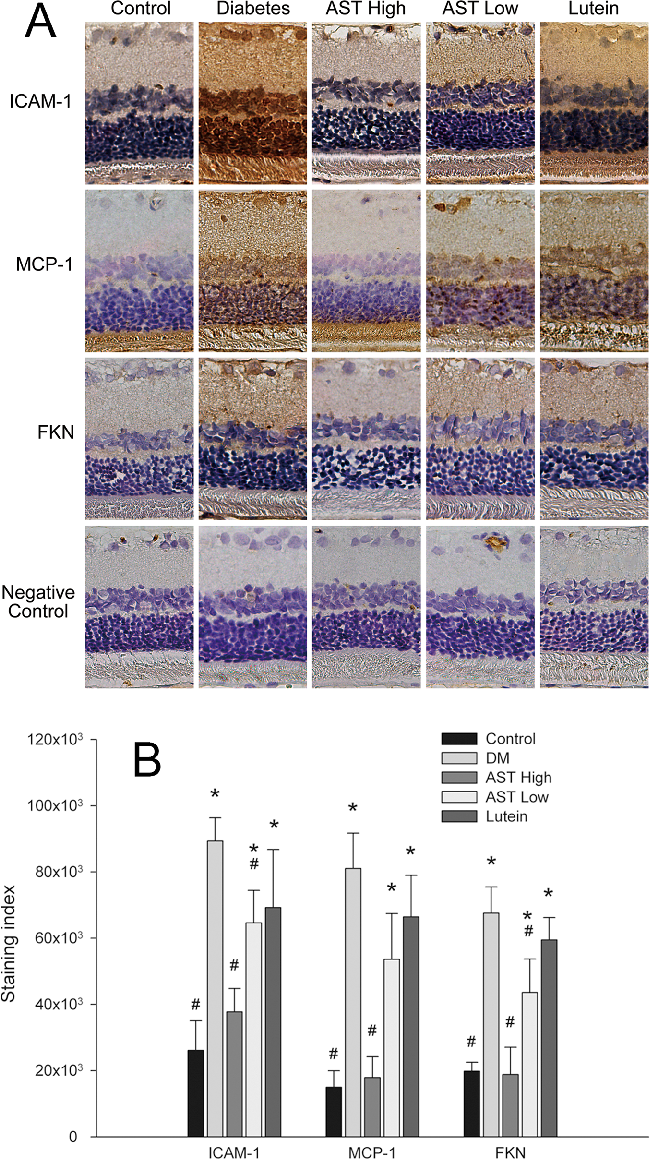
Histological or immunohistochemical analysis of retinal tissue sections from diabetic rats, comparing structural changes across treatment groups to assess astaxanthin's protective effects on retinal architecture.
Astaxanthin Inhibits Expression of Retinal Oxidative Stress and Inflammatory Mediators in Streptozotocin-Induced …
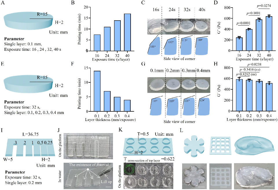
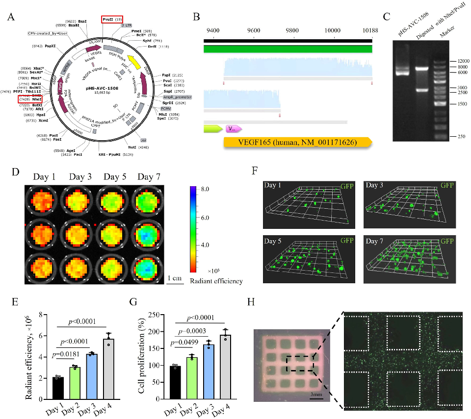
VEGF165-overexpressing endothelial cells encapsulated within the hydrogel are assessed for viability and vascular network formation. The growth factor expression aims to promote angiogenesis, addressing the vascular injury central to non-healing diabetic wounds.
A click chemistry-mediated all-peptide cell printing hydrogel platform for diabetic wound healing.
In vitro wound healing assays using the bioprinted hydrogel constructs demonstrate enhanced cell migration and proliferation. The results suggest that the all-peptide platform supports the cellular processes required for tissue regeneration.
A click chemistry-mediated all-peptide cell printing hydrogel platform for diabetic wound healing.
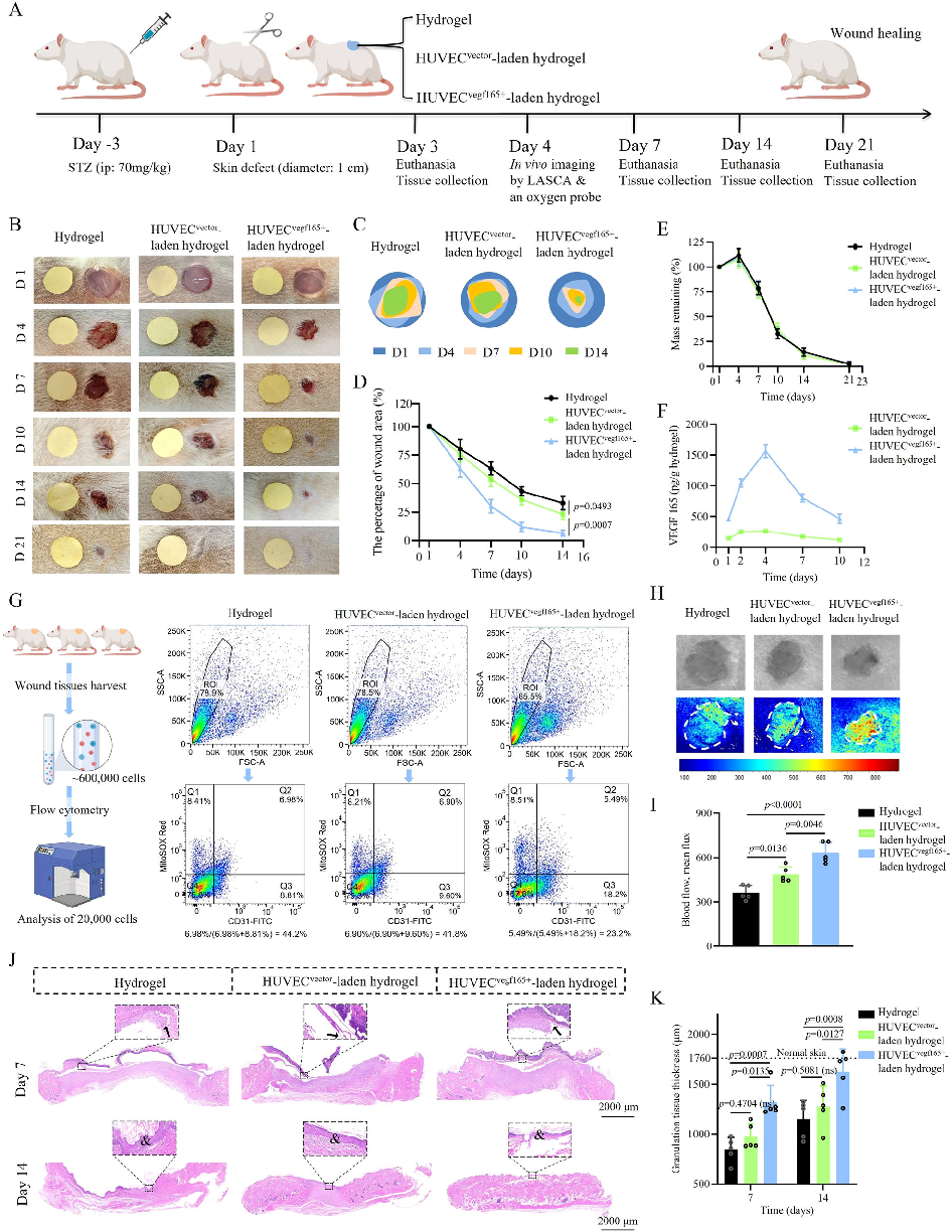
Histological analysis of healed wound tissue reveals improved tissue architecture and vascularization in the hydrogel-treated group. Hematoxylin and eosin staining shows more organized collagen deposition and reduced inflammatory infiltrate.
A click chemistry-mediated all-peptide cell printing hydrogel platform for diabetic wound healing.
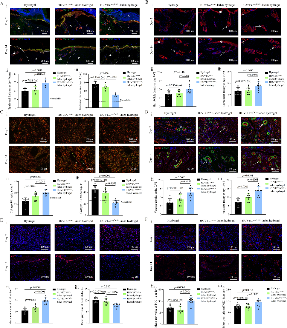
Immunohistochemical staining of wound sections confirms enhanced angiogenesis in hydrogel-treated diabetic wounds. Markers for vascular endothelial cells and smooth muscle cells indicate formation of functional blood vessels in the regenerated tissue.
A click chemistry-mediated all-peptide cell printing hydrogel platform for diabetic wound healing.
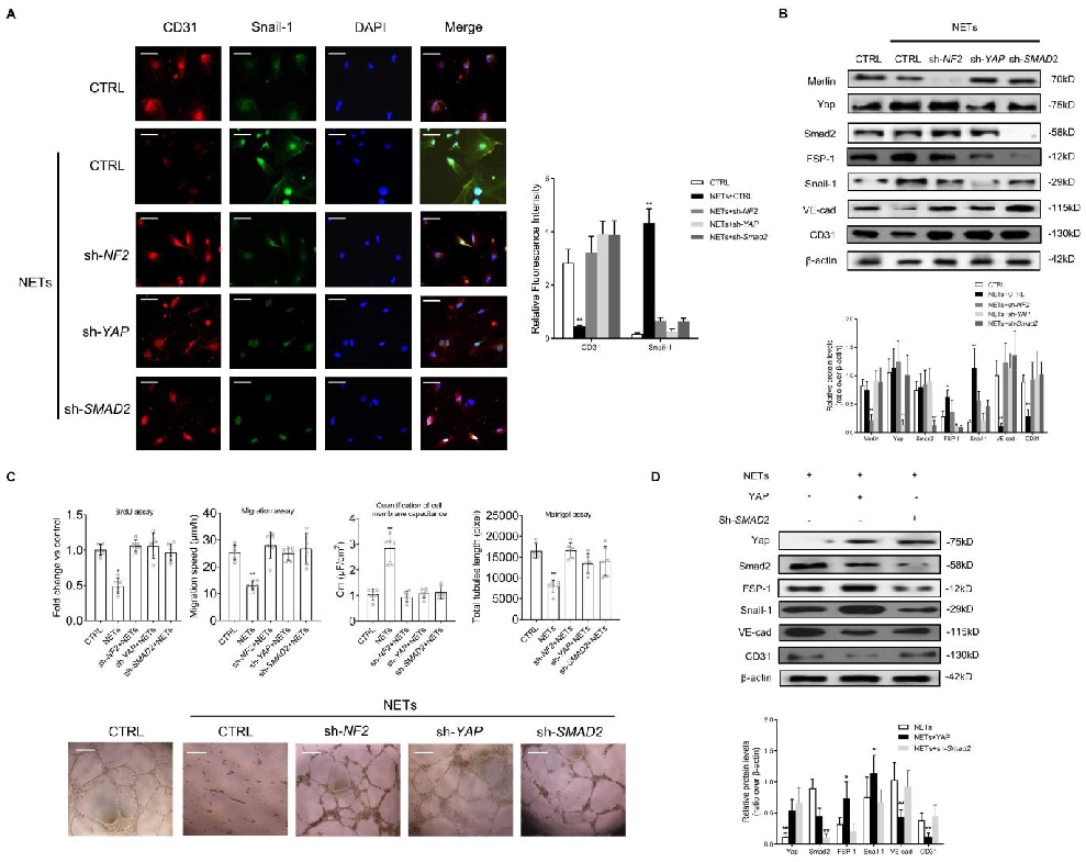
Histological analysis of wound tissue from NET-depleted and control diabetic mice reveals improved re-epithelialization and vascularization.
Neutrophil Extracellular Traps Delay Diabetic Wound Healing by Inducing Endothelial-to-Mesenchymal Transition via …