Arginine 图表
38 来自同行评审研究的图表
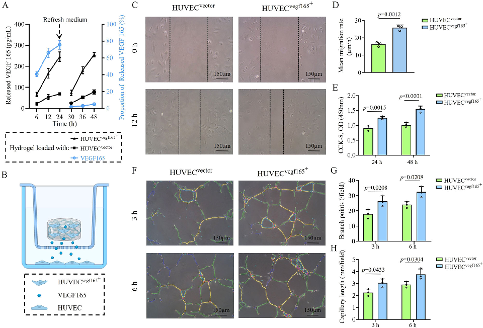
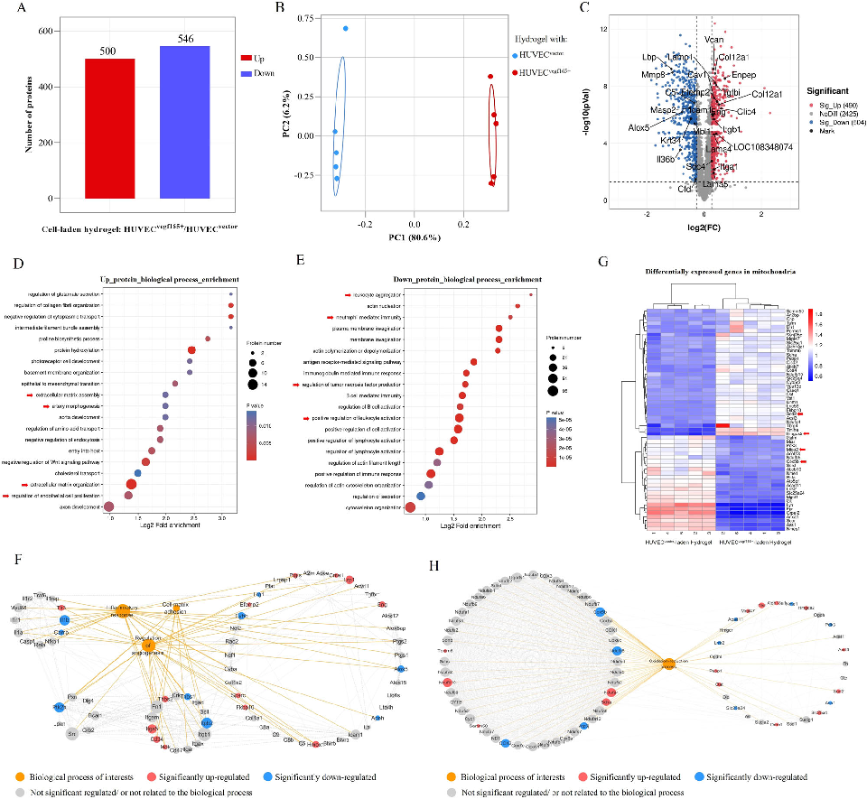
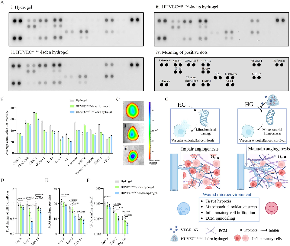
Gene expression or protein analysis from cells cultured in the hydrogel platform reveals upregulation of angiogenic and wound healing markers. The molecular data support the functional benefits observed in cell migration and proliferation assays.
A click chemistry-mediated all-peptide cell printing hydrogel platform for diabetic wound healing.
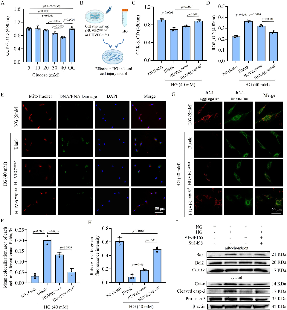
In vivo evaluation of the bioprinted hydrogel in a diabetic wound model shows wound closure progression over time. The cell-laden constructs demonstrate accelerated healing compared to control treatments in the high-glucose wound environment.
A click chemistry-mediated all-peptide cell printing hydrogel platform for diabetic wound healing.
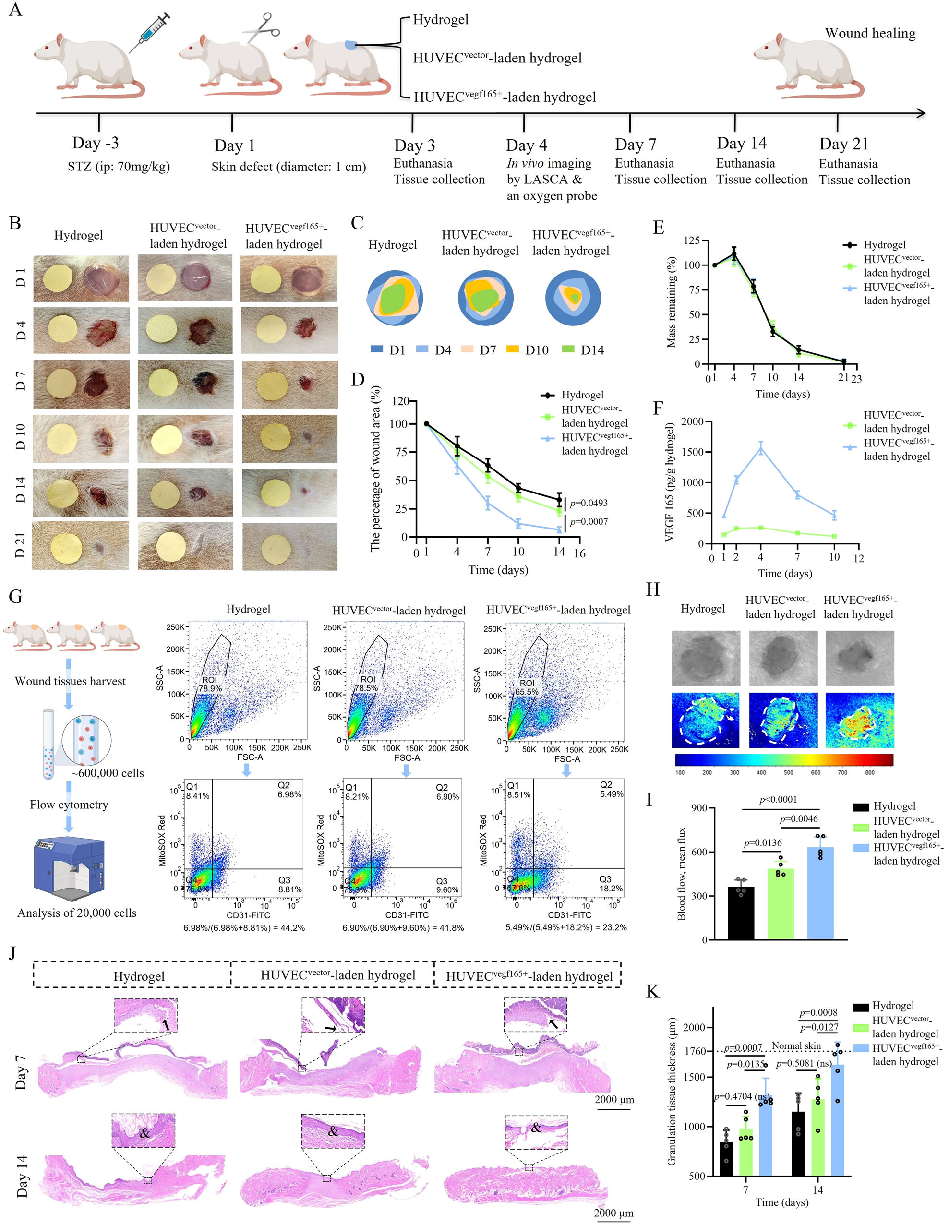
Histological analysis of healed wound tissue reveals improved tissue architecture and vascularization in the hydrogel-treated group. Hematoxylin and eosin staining shows more organized collagen deposition and reduced inflammatory infiltrate.
A click chemistry-mediated all-peptide cell printing hydrogel platform for diabetic wound healing.
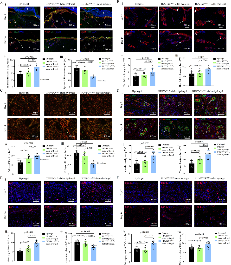
Immunohistochemical staining of wound sections confirms enhanced angiogenesis in hydrogel-treated diabetic wounds. Markers for vascular endothelial cells and smooth muscle cells indicate formation of functional blood vessels in the regenerated tissue.
A click chemistry-mediated all-peptide cell printing hydrogel platform for diabetic wound healing.
Quantitative analysis of wound healing outcomes compares the bioprinted hydrogel against conventional treatments. Metrics including wound closure rate, collagen density, and vessel density are significantly improved in the treatment group.
A click chemistry-mediated all-peptide cell printing hydrogel platform for diabetic wound healing.
Supplementary characterization or additional in vivo data from the click chemistry hydrogel study is presented. The comprehensive evaluation supports the platform's potential for translational application in diabetic wound management.
A click chemistry-mediated all-peptide cell printing hydrogel platform for diabetic wound healing.
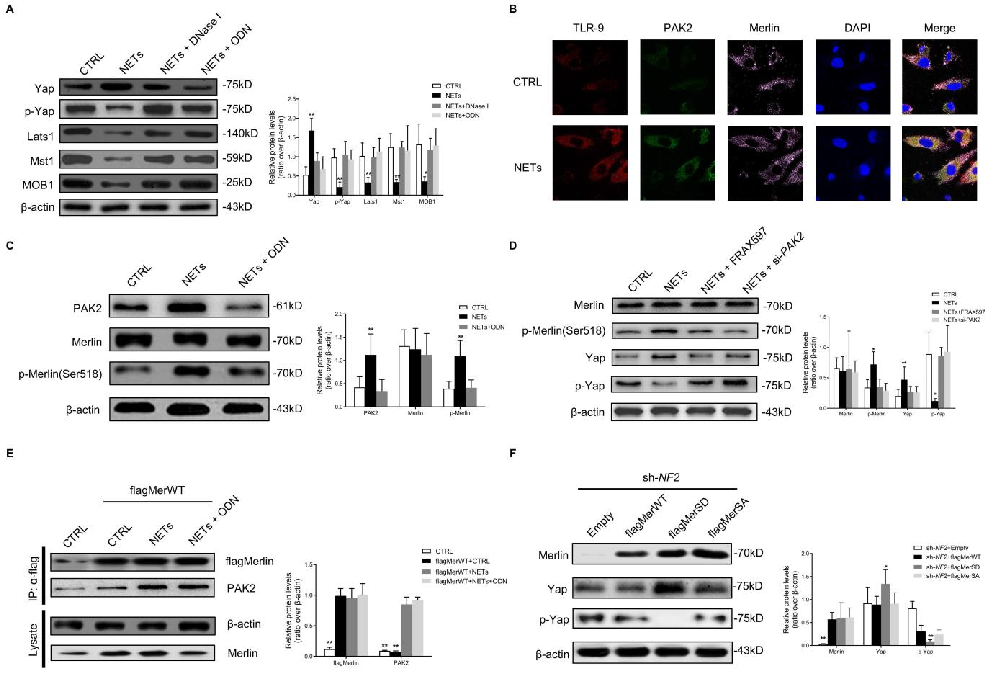
The molecular mechanism linking NET-derived DNA to Hippo-YAP pathway inhibition and endothelial-to-mesenchymal transition is elucidated, with DNase treatment as a potential therapeutic approach.
Neutrophil Extracellular Traps Delay Diabetic Wound Healing by Inducing Endothelial-to-Mesenchymal Transition via …
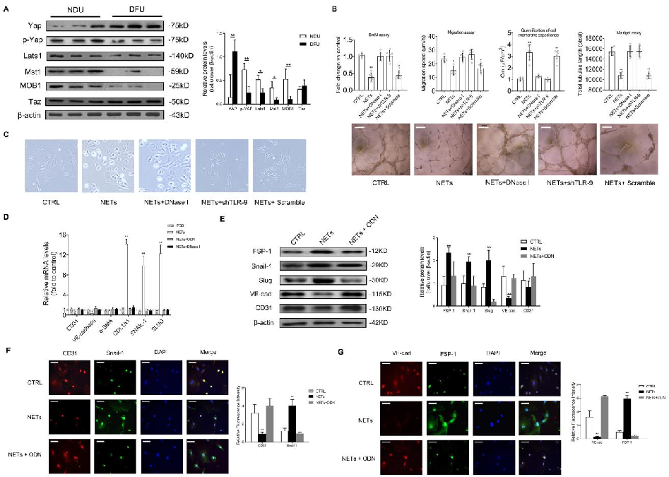
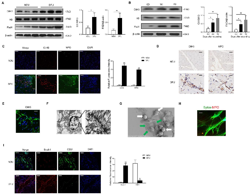
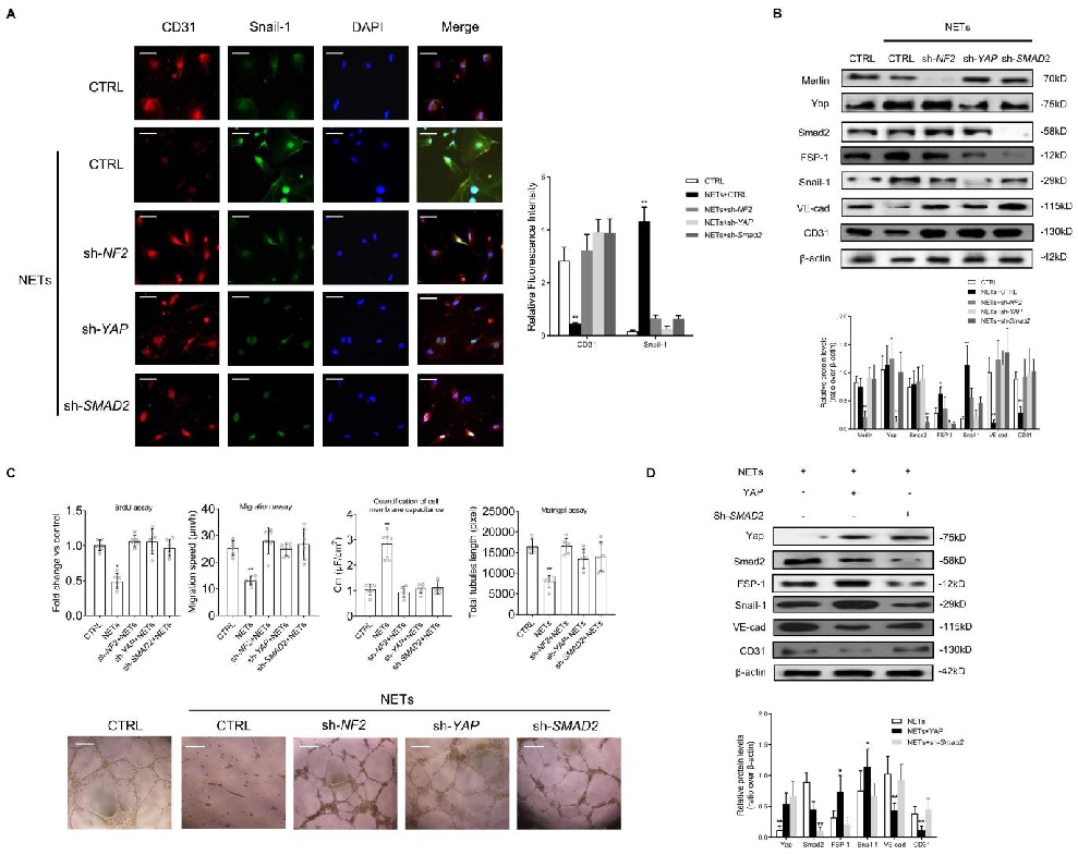
In vitro endothelial-to-mesenchymal transition markers are quantified following NET exposure, demonstrating increased mesenchymal and decreased endothelial protein expression.
Neutrophil Extracellular Traps Delay Diabetic Wound Healing by Inducing Endothelial-to-Mesenchymal Transition via …
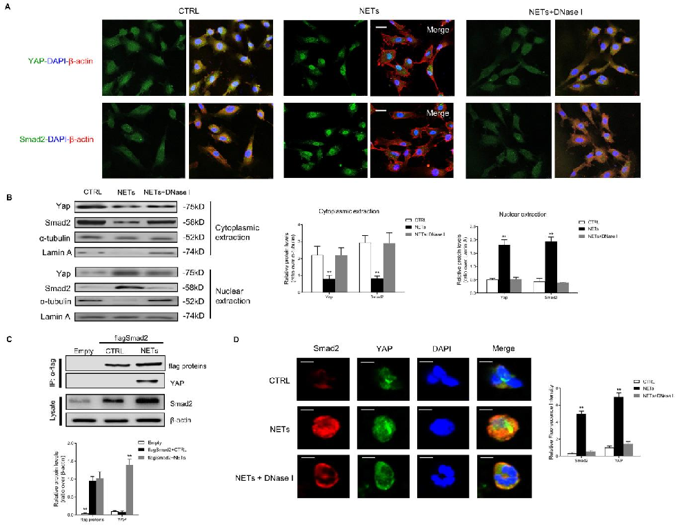
YAP phosphorylation and nuclear exclusion in NET-treated endothelial cells are characterized, linking NET exposure to Hippo pathway activation.
Neutrophil Extracellular Traps Delay Diabetic Wound Healing by Inducing Endothelial-to-Mesenchymal Transition via …
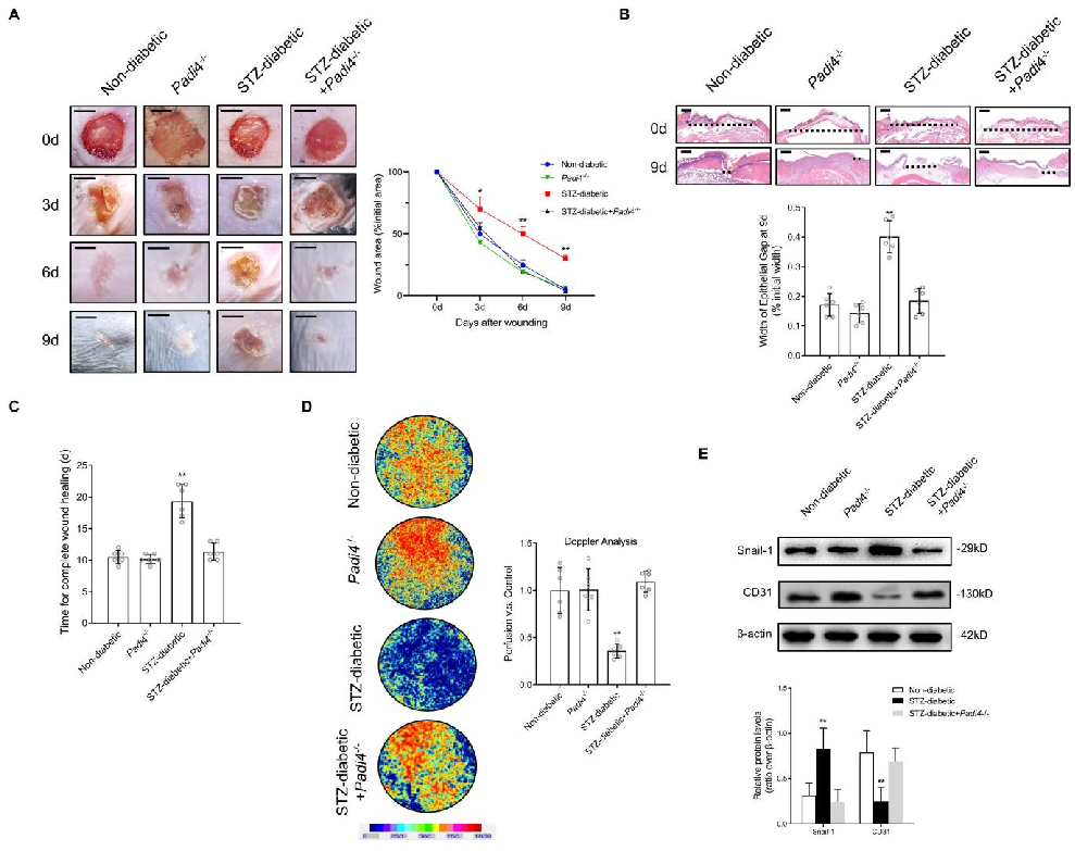
Wound healing outcomes in Padi4-knockout diabetic mice compared to wild-type controls are documented, showing accelerated closure when NET formation is genetically ablated.
Neutrophil Extracellular Traps Delay Diabetic Wound Healing by Inducing Endothelial-to-Mesenchymal Transition via …
Histological analysis of wound tissue from NET-depleted and control diabetic mice reveals improved re-epithelialization and vascularization.
Neutrophil Extracellular Traps Delay Diabetic Wound Healing by Inducing Endothelial-to-Mesenchymal Transition via …
DNase I treatment effects on diabetic wound healing in vivo are quantified, demonstrating that NET degradation promotes wound closure through preserved endothelial function.
Neutrophil Extracellular Traps Delay Diabetic Wound Healing by Inducing Endothelial-to-Mesenchymal Transition via …
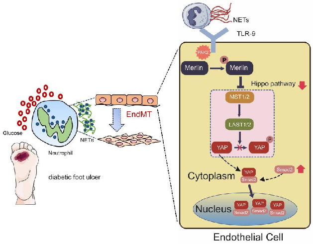
A proposed model integrating NET-mediated Hippo pathway suppression with endothelial-to-mesenchymal transition in delayed diabetic wound healing is diagrammed.
Neutrophil Extracellular Traps Delay Diabetic Wound Healing by Inducing Endothelial-to-Mesenchymal Transition via …
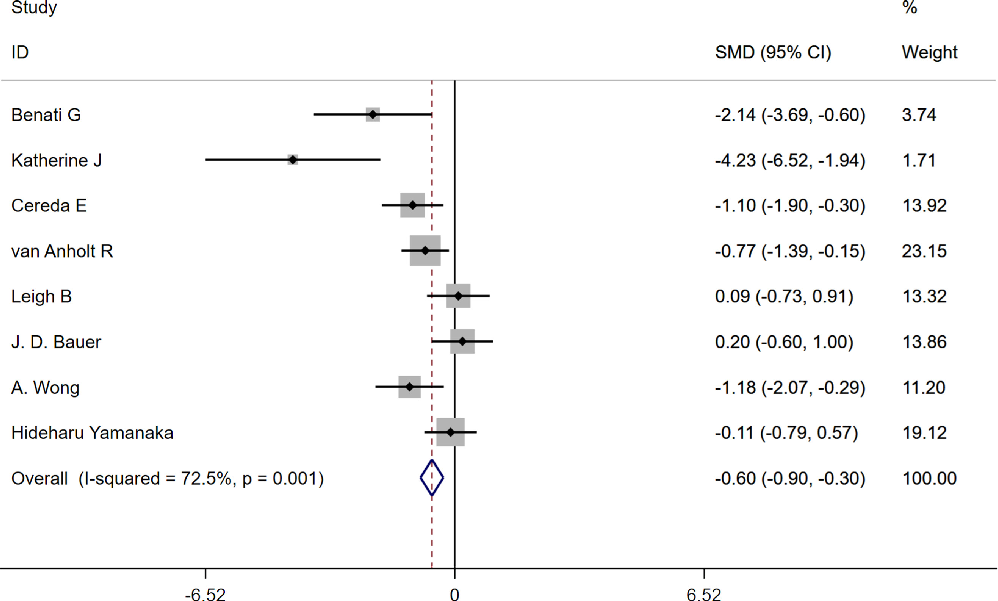
Conceptual diagram summarizing the relationship between the use of oral and enteral tube and the biological processes described in this research.
The use of oral and enteral tube-fed arginine supplementation in pressure injury …
第 2 页,共 2 页