Forschungsprozess
333 Abbildungen aus begutachteter Forschung
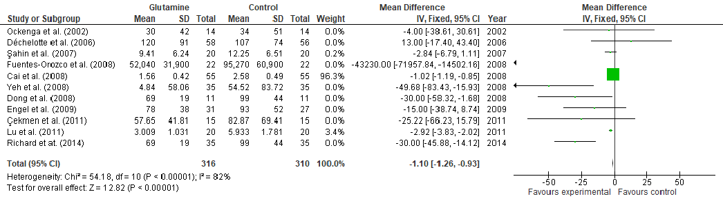
C-reactive protein levels following glutamine supplementation are pooled in a fixed-effects forest plot. Lower CRP values suggest reduced systemic inflammation, which may contribute to improved wound healing outcomes.
The Effect of Amino Acids on Wound Healing: A Systematic Review and …
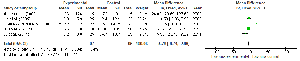
An additional inflammatory or immune marker from the glutamine meta-analysis is displayed in forest plot format. The pooled analysis evaluates whether glutamine supplementation modulates immune parameters relevant to wound repair.
The Effect of Amino Acids on Wound Healing: A Systematic Review and …
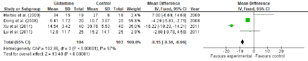
Further meta-analysis results present pooled effect sizes for wound healing parameters in glutamine-supplemented patients. The forest plot format allows visual comparison of effect magnitude and consistency across trials.
The Effect of Amino Acids on Wound Healing: A Systematic Review and …
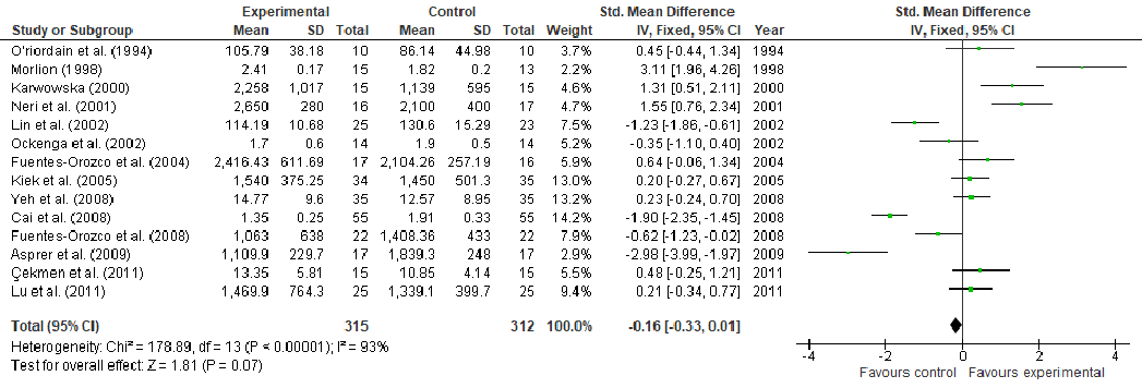
T-cell lymphocyte counts are analyzed in a fixed-effects forest plot across glutamine supplementation trials. Enhanced T-cell proliferation is associated with improved immune-mediated wound repair, and the pooled estimate evaluates glutamine's immunomodulatory potential.
The Effect of Amino Acids on Wound Healing: A Systematic Review and …
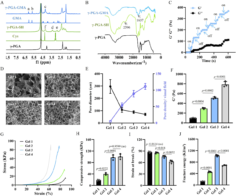
Rheological and mechanical characterization of the click chemistry hydrogel demonstrates properties suitable for 3D bioprinting. The material's shear-thinning behavior and rapid recovery enable precise deposition of cell-laden constructs.
A click chemistry-mediated all-peptide cell printing hydrogel platform for diabetic wound healing.
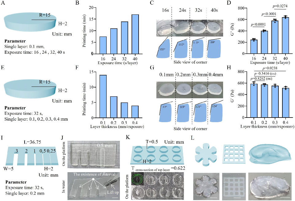
Bioprinting parameters and construct fidelity are evaluated for the peptide hydrogel system. The printability assessment confirms that the material maintains structural integrity while supporting embedded vascular endothelial cell viability.
A click chemistry-mediated all-peptide cell printing hydrogel platform for diabetic wound healing.
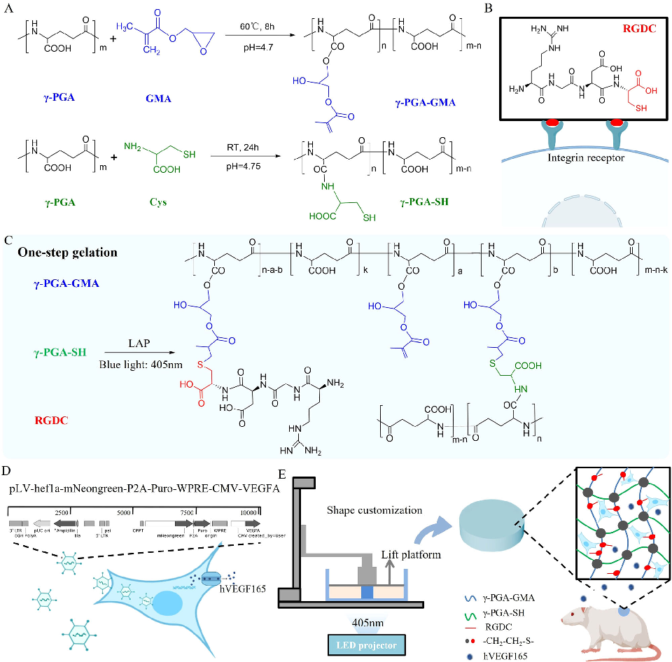
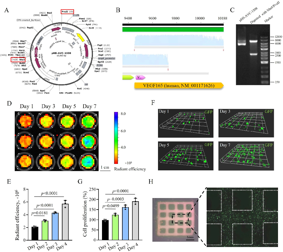
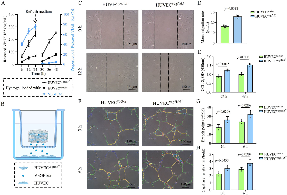
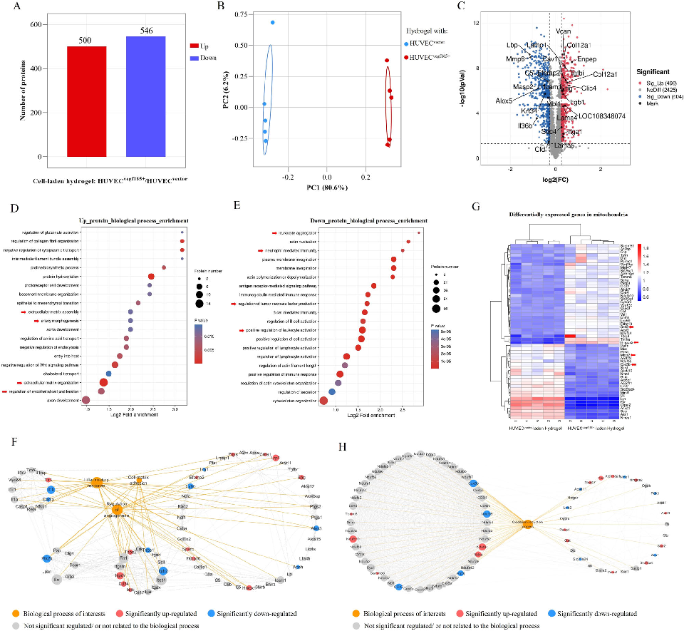
VEGF165-overexpressing endothelial cells encapsulated within the hydrogel are assessed for viability and vascular network formation. The growth factor expression aims to promote angiogenesis, addressing the vascular injury central to non-healing diabetic wounds.
A click chemistry-mediated all-peptide cell printing hydrogel platform for diabetic wound healing.
In vitro wound healing assays using the bioprinted hydrogel constructs demonstrate enhanced cell migration and proliferation. The results suggest that the all-peptide platform supports the cellular processes required for tissue regeneration.
A click chemistry-mediated all-peptide cell printing hydrogel platform for diabetic wound healing.
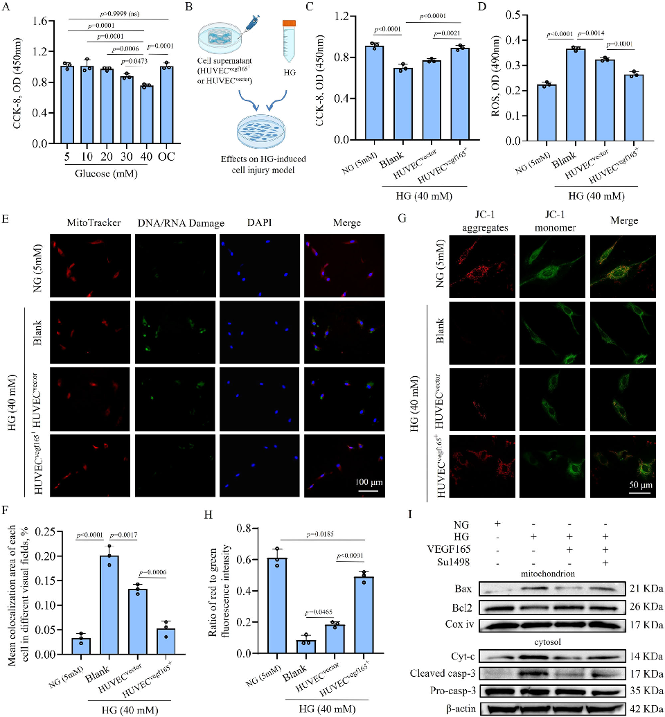
Gene expression or protein analysis from cells cultured in the hydrogel platform reveals upregulation of angiogenic and wound healing markers. The molecular data support the functional benefits observed in cell migration and proliferation assays.
A click chemistry-mediated all-peptide cell printing hydrogel platform for diabetic wound healing.
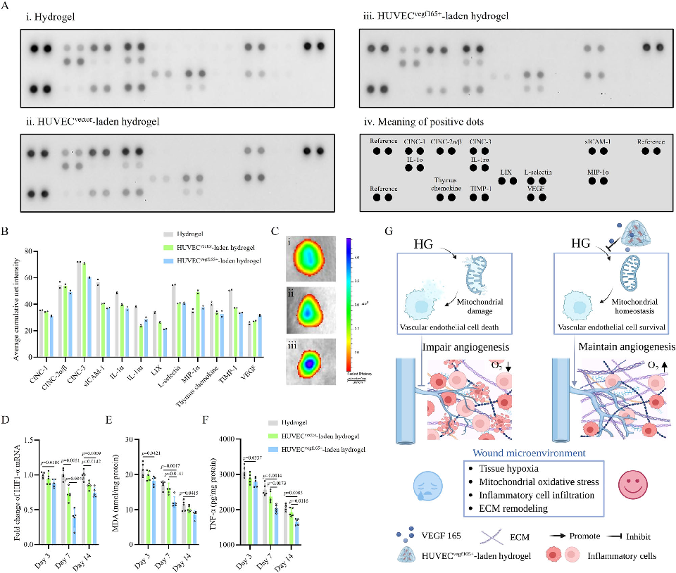
In vivo evaluation of the bioprinted hydrogel in a diabetic wound model shows wound closure progression over time. The cell-laden constructs demonstrate accelerated healing compared to control treatments in the high-glucose wound environment.
A click chemistry-mediated all-peptide cell printing hydrogel platform for diabetic wound healing.
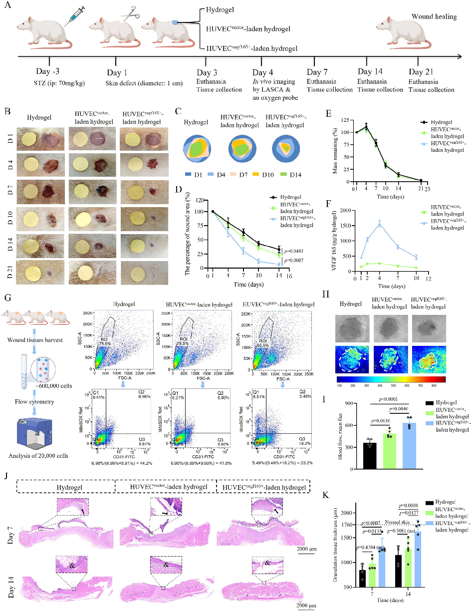
Histological analysis of healed wound tissue reveals improved tissue architecture and vascularization in the hydrogel-treated group. Hematoxylin and eosin staining shows more organized collagen deposition and reduced inflammatory infiltrate.
A click chemistry-mediated all-peptide cell printing hydrogel platform for diabetic wound healing.
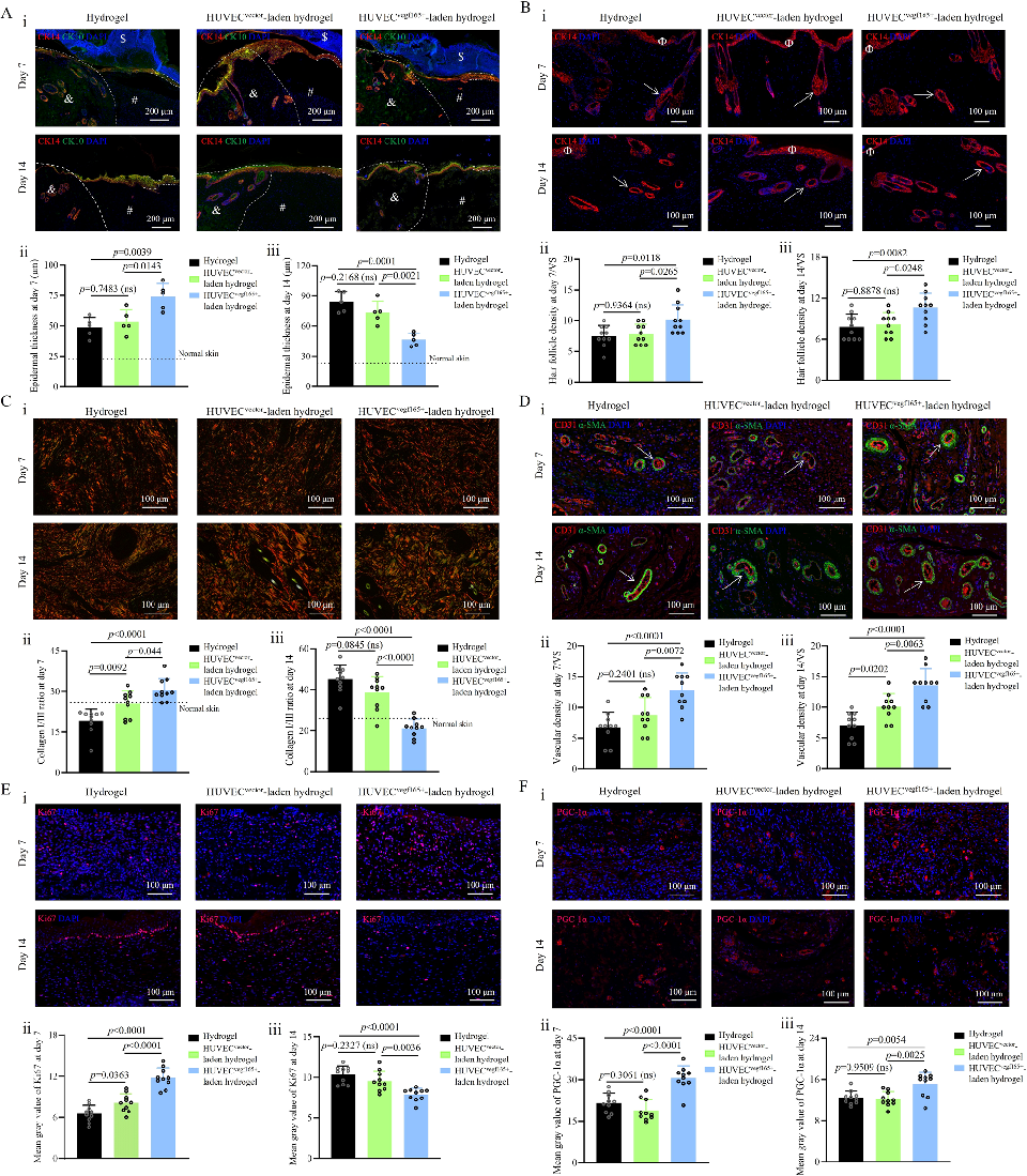
Immunohistochemical staining of wound sections confirms enhanced angiogenesis in hydrogel-treated diabetic wounds. Markers for vascular endothelial cells and smooth muscle cells indicate formation of functional blood vessels in the regenerated tissue.
A click chemistry-mediated all-peptide cell printing hydrogel platform for diabetic wound healing.
Quantitative analysis of wound healing outcomes compares the bioprinted hydrogel against conventional treatments. Metrics including wound closure rate, collagen density, and vessel density are significantly improved in the treatment group.
A click chemistry-mediated all-peptide cell printing hydrogel platform for diabetic wound healing.
Supplementary characterization or additional in vivo data from the click chemistry hydrogel study is presented. The comprehensive evaluation supports the platform's potential for translational application in diabetic wound management.
A click chemistry-mediated all-peptide cell printing hydrogel platform for diabetic wound healing.
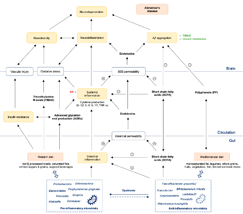
A pathway diagram illustrates how gut dysbiosis promotes intestinal and systemic inflammation, leading to amyloid-beta aggregation, neuroinflammation, and ultimately neurodegeneration in Alzheimer's disease. The cascade connects microbial imbalance to blood-brain barrier compromise and central nervous system pathology.
The Immunopathogenesis of Alzheimer's Disease Is Related to the Composition of Gut …
Western diet-induced microbiome changes are mapped to intestinal dysbiosis, low-grade gut inflammation, and increased permeability of both the intestinal barrier and blood-brain barrier. The resulting systemic inflammatory state is linked to neuroinflammatory processes implicated in Alzheimer's disease progression.
The Immunopathogenesis of Alzheimer's Disease Is Related to the Composition of Gut …
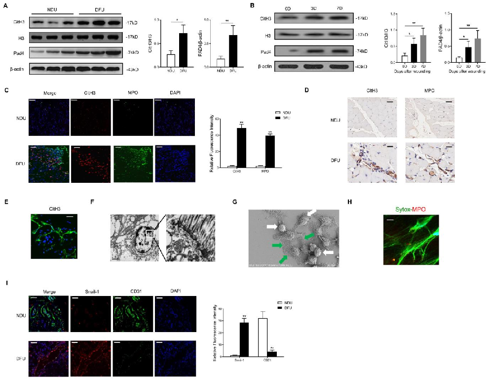
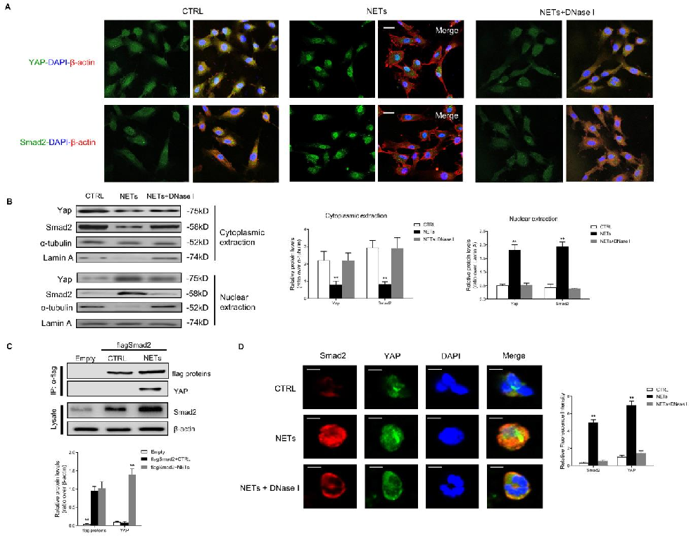
The molecular mechanism linking NET-derived DNA to Hippo-YAP pathway inhibition and endothelial-to-mesenchymal transition is elucidated, with DNase treatment as a potential therapeutic approach.
Neutrophil Extracellular Traps Delay Diabetic Wound Healing by Inducing Endothelial-to-Mesenchymal Transition via …
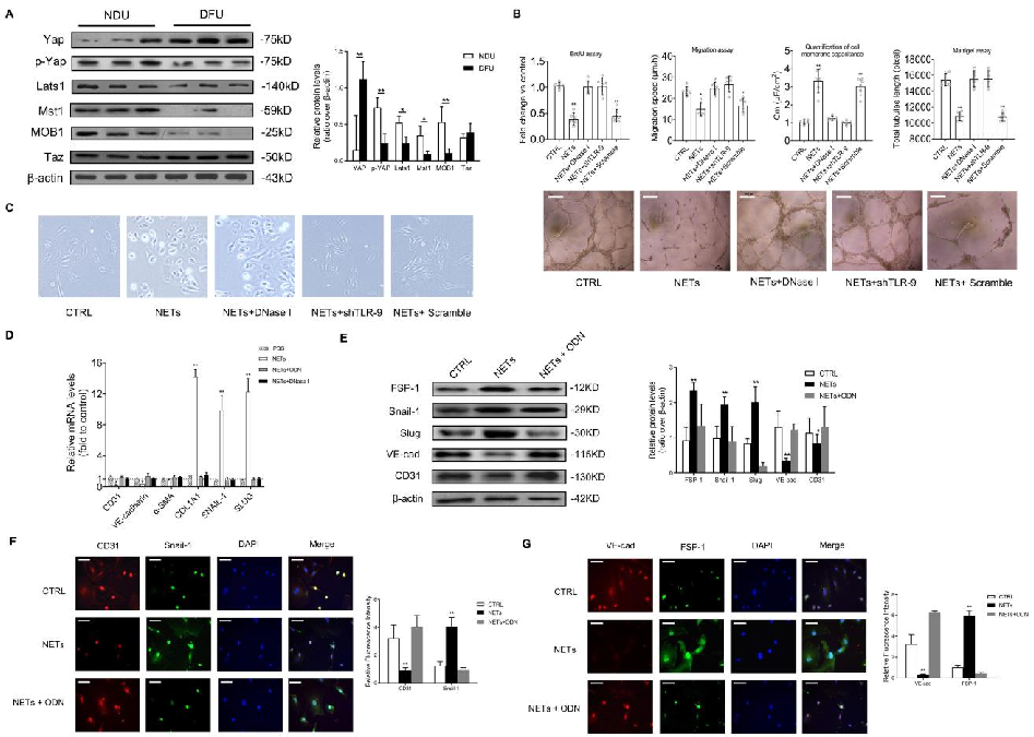
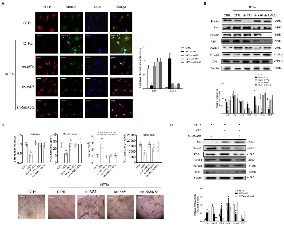
In vitro endothelial-to-mesenchymal transition markers are quantified following NET exposure, demonstrating increased mesenchymal and decreased endothelial protein expression.
Neutrophil Extracellular Traps Delay Diabetic Wound Healing by Inducing Endothelial-to-Mesenchymal Transition via …
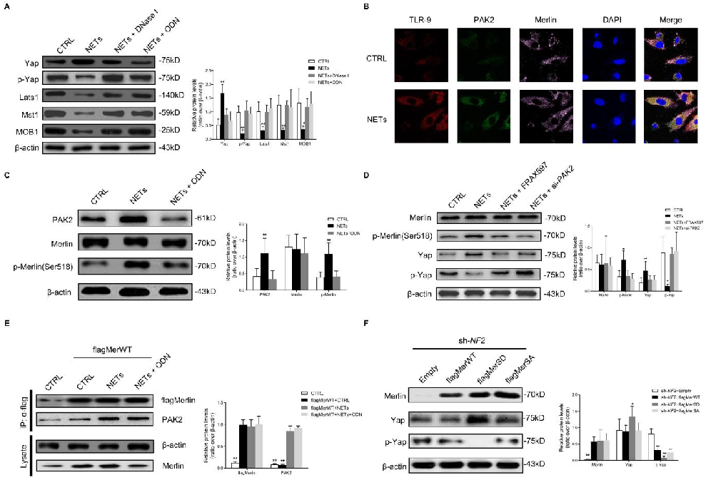
YAP phosphorylation and nuclear exclusion in NET-treated endothelial cells are characterized, linking NET exposure to Hippo pathway activation.
Neutrophil Extracellular Traps Delay Diabetic Wound Healing by Inducing Endothelial-to-Mesenchymal Transition via …
Wound healing outcomes in Padi4-knockout diabetic mice compared to wild-type controls are documented, showing accelerated closure when NET formation is genetically ablated.
Neutrophil Extracellular Traps Delay Diabetic Wound Healing by Inducing Endothelial-to-Mesenchymal Transition via …
Histological analysis of wound tissue from NET-depleted and control diabetic mice reveals improved re-epithelialization and vascularization.
Neutrophil Extracellular Traps Delay Diabetic Wound Healing by Inducing Endothelial-to-Mesenchymal Transition via …
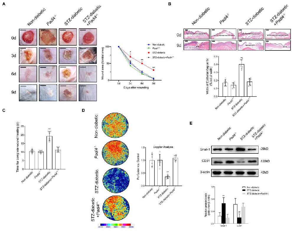
DNase I treatment effects on diabetic wound healing in vivo are quantified, demonstrating that NET degradation promotes wound closure through preserved endothelial function.
Neutrophil Extracellular Traps Delay Diabetic Wound Healing by Inducing Endothelial-to-Mesenchymal Transition via …
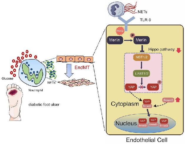
A proposed model integrating NET-mediated Hippo pathway suppression with endothelial-to-mesenchymal transition in delayed diabetic wound healing is diagrammed.
Neutrophil Extracellular Traps Delay Diabetic Wound Healing by Inducing Endothelial-to-Mesenchymal Transition via …
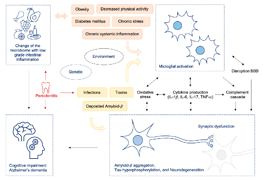
Brain insulin resistance mechanisms underlying depression are depicted, showing how HPA axis dysregulation, reduced anterior cingulate cortex volume, and impaired hippocampal function contribute to the condition.
A review on linking stress, depression, and insulin resistance via low-grade chronic …
Seite 5 von 14