Arginine Figure
11 figure da ricerca revisionata da esperti
Risk of bias assessment for arginine studies is displayed using the Cochrane tool framework. Each domain of potential bias is evaluated across the included trials to gauge the overall quality of evidence supporting arginine supplementation for wound healing.
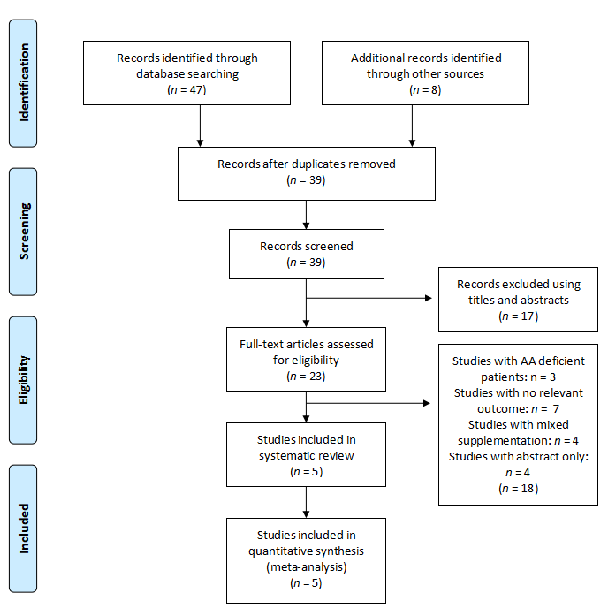
The Effect of Amino Acids on Wound Healing: A Systematic Review and …
Risk of bias assessment for glutamine studies complements the arginine evaluation. The systematic assessment of selection, performance, detection, attrition, and reporting bias helps contextualize the strength of conclusions about glutamine and wound repair.
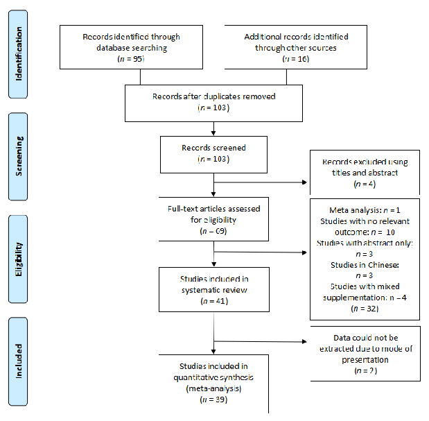
The Effect of Amino Acids on Wound Healing: A Systematic Review and …
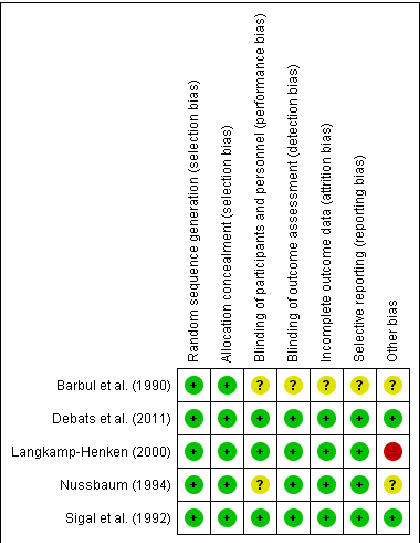
Risk of bias is summarized for each included arginine study using green (low risk), yellow (unclear), and red (high risk) indicators. The visual matrix helps readers quickly assess methodological quality across randomization, blinding, and outcome reporting domains.
The Effect of Amino Acids on Wound Healing: A Systematic Review and …
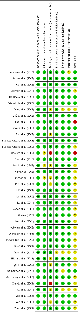
Risk of bias for the included glutamine studies follows the same Cochrane framework, with symbols indicating low, unclear, and high risk across multiple domains. The assessment covers 39 human studies evaluating glutamine supplementation effects on wound healing.
The Effect of Amino Acids on Wound Healing: A Systematic Review and …
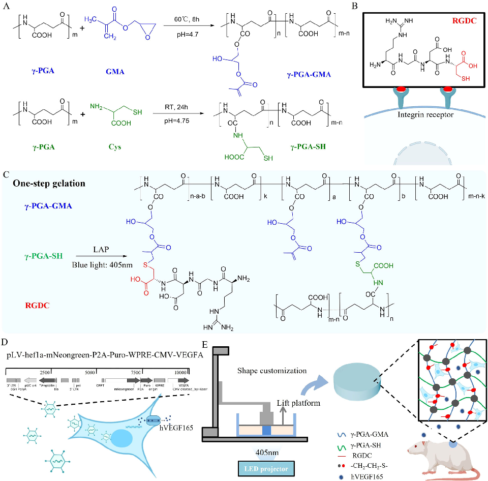
Rheological and mechanical characterization of the click chemistry hydrogel demonstrates properties suitable for 3D bioprinting. The material's shear-thinning behavior and rapid recovery enable precise deposition of cell-laden constructs.
A click chemistry-mediated all-peptide cell printing hydrogel platform for diabetic wound healing.
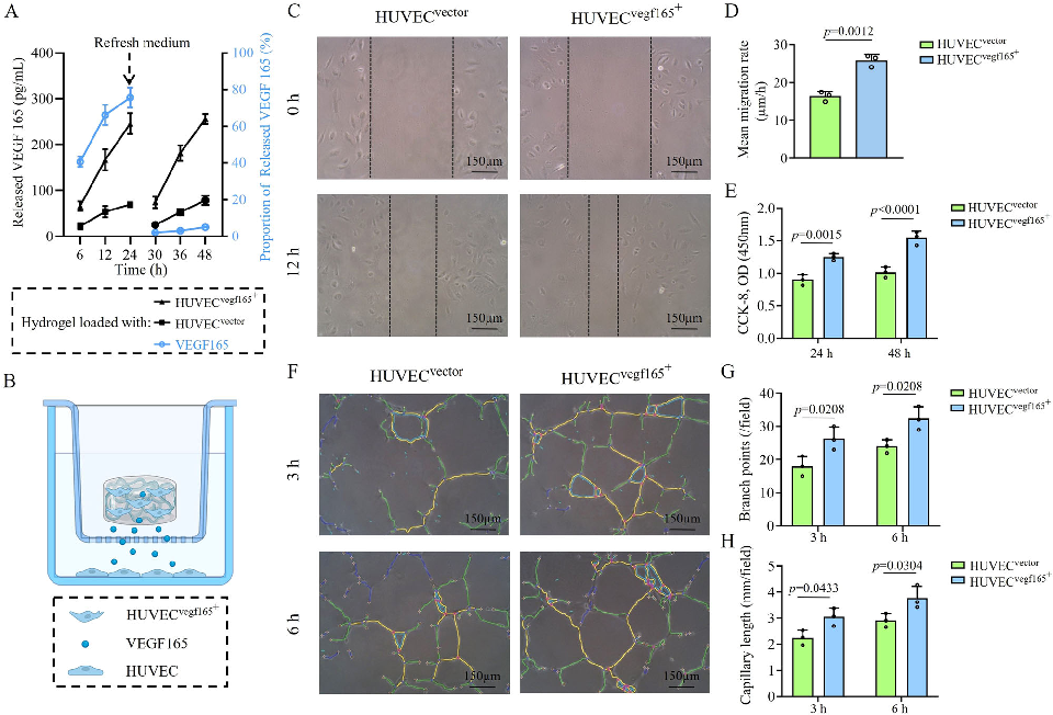
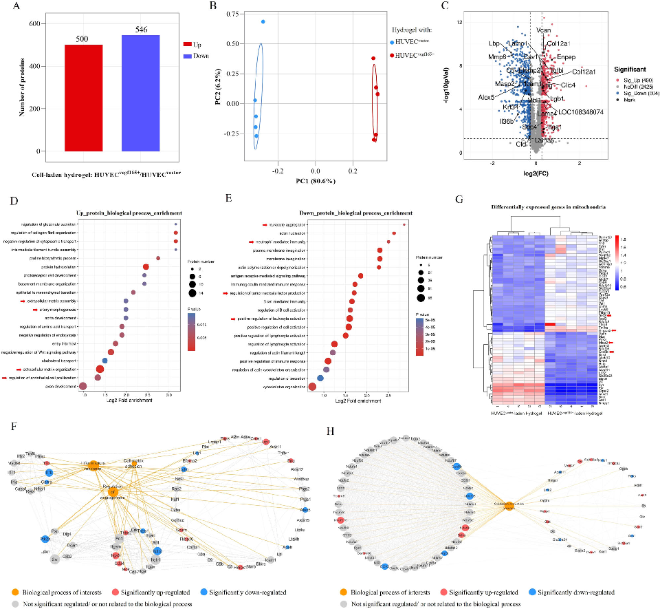
Gene expression or protein analysis from cells cultured in the hydrogel platform reveals upregulation of angiogenic and wound healing markers. The molecular data support the functional benefits observed in cell migration and proliferation assays.
A click chemistry-mediated all-peptide cell printing hydrogel platform for diabetic wound healing.
Quantitative analysis of wound healing outcomes compares the bioprinted hydrogel against conventional treatments. Metrics including wound closure rate, collagen density, and vessel density are significantly improved in the treatment group.
A click chemistry-mediated all-peptide cell printing hydrogel platform for diabetic wound healing.
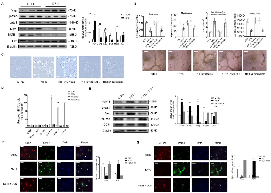
In vitro endothelial-to-mesenchymal transition markers are quantified following NET exposure, demonstrating increased mesenchymal and decreased endothelial protein expression.
Neutrophil Extracellular Traps Delay Diabetic Wound Healing by Inducing Endothelial-to-Mesenchymal Transition via …
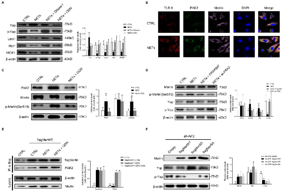
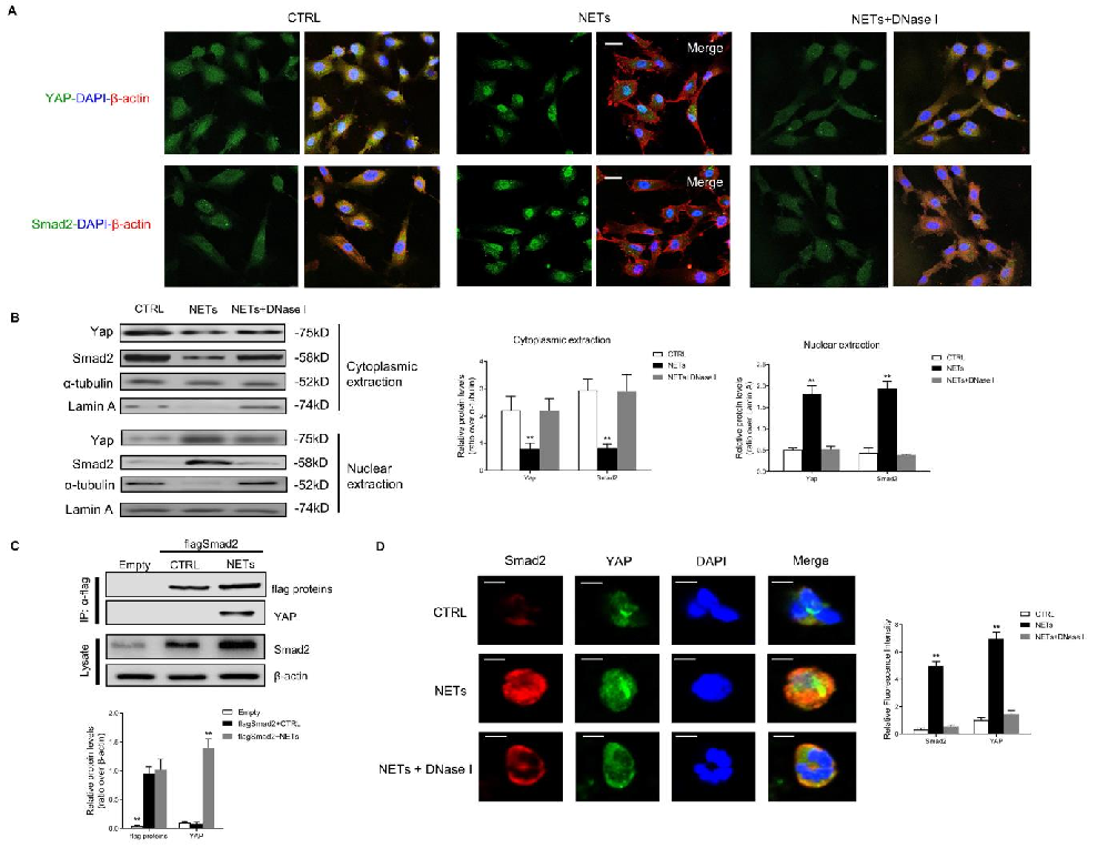
YAP phosphorylation and nuclear exclusion in NET-treated endothelial cells are characterized, linking NET exposure to Hippo pathway activation.
Neutrophil Extracellular Traps Delay Diabetic Wound Healing by Inducing Endothelial-to-Mesenchymal Transition via …
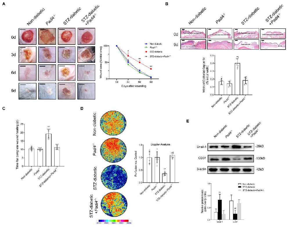
Wound healing outcomes in Padi4-knockout diabetic mice compared to wild-type controls are documented, showing accelerated closure when NET formation is genetically ablated.
Neutrophil Extracellular Traps Delay Diabetic Wound Healing by Inducing Endothelial-to-Mesenchymal Transition via …
DNase I treatment effects on diabetic wound healing in vivo are quantified, demonstrating that NET degradation promotes wound closure through preserved endothelial function.
Neutrophil Extracellular Traps Delay Diabetic Wound Healing by Inducing Endothelial-to-Mesenchymal Transition via …