Arginine Figure
6 figure da ricerca revisionata da esperti
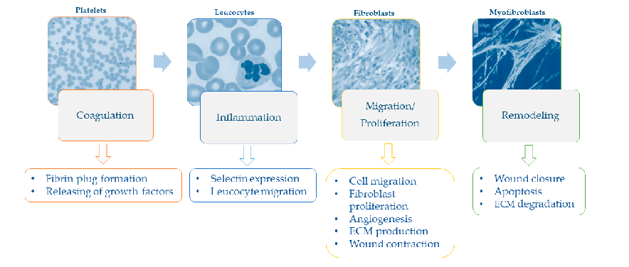
Sequential phases of the wound healing process - hemostasis, inflammation, proliferation, and remodeling - and their specific cellular events are illustrated.
Nutrition and Wound Healing: An Overview Focusing on the Beneficial Effects of …
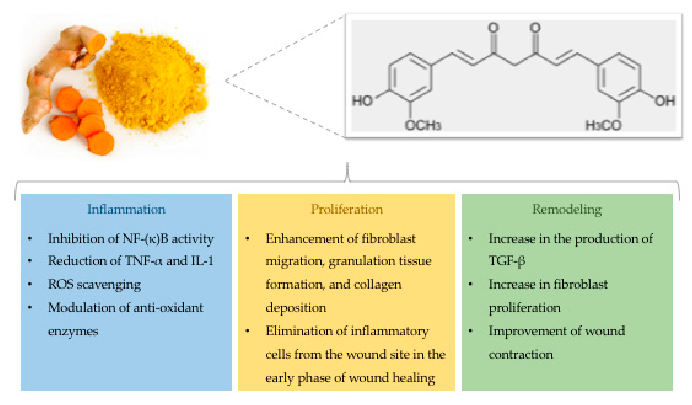
Chemical structure of curcumin alongside its documented effects on wound healing, including anti-inflammatory, antioxidant, and antimicrobial activities, are presented.
Nutrition and Wound Healing: An Overview Focusing on the Beneficial Effects of …
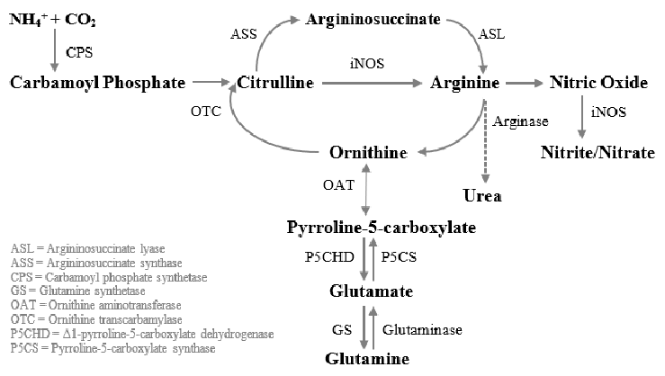
Glutamine-to-arginine metabolism in human macrophages is mapped, showing the conversion of carbamoyl phosphate and ornithine to citrulline via ornithine transcarbamylase, and subsequent transformation to argininosuccinate. This pathway is central to understanding how glutamine supports immune-mediated wound repair.
The Effect of Amino Acids on Wound Healing: A Systematic Review and …
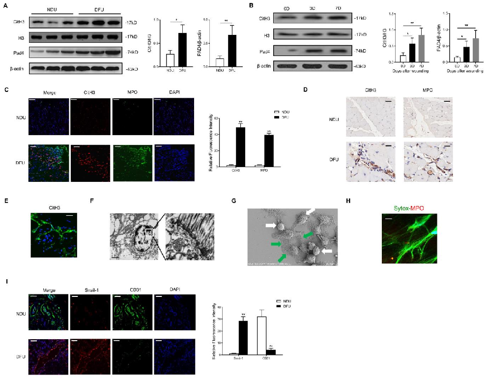
The molecular mechanism linking NET-derived DNA to Hippo-YAP pathway inhibition and endothelial-to-mesenchymal transition is elucidated, with DNase treatment as a potential therapeutic approach.
Neutrophil Extracellular Traps Delay Diabetic Wound Healing by Inducing Endothelial-to-Mesenchymal Transition via …
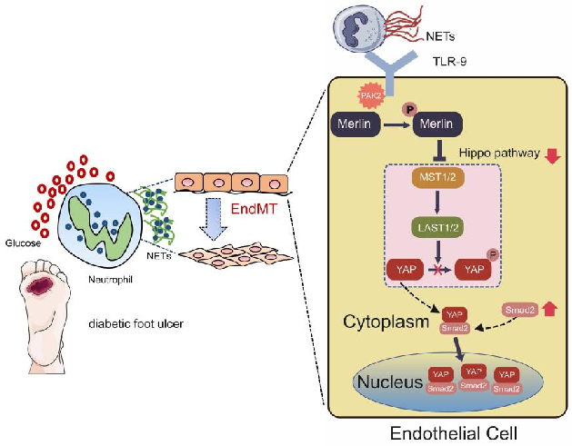
A proposed model integrating NET-mediated Hippo pathway suppression with endothelial-to-mesenchymal transition in delayed diabetic wound healing is diagrammed.
Neutrophil Extracellular Traps Delay Diabetic Wound Healing by Inducing Endothelial-to-Mesenchymal Transition via …
Conceptual diagram summarizing the relationship between the use of oral and enteral tube and the biological processes described in this research.
The use of oral and enteral tube-fed arginine supplementation in pressure injury …