研究流程
333 来自同行评审研究的图表
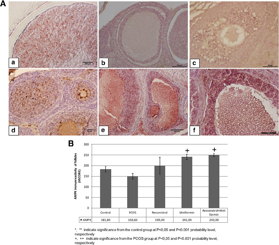
AMPK immunoreactivity analysis reveals decreased phosphorylated AMPK in PCOS ovarian tissue compared to controls. Both metformin and resveratrol treatments partially restore AMPK activation, consistent with their known metabolic signaling effects.
Effect of resveratrol and metformin on ovarian reserve and ultrastructure in PCOS: …
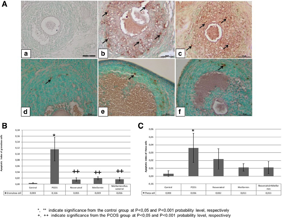
TUNEL analysis of granulosa and theca cells quantifies apoptotic rates across experimental groups. PCOS induction increases apoptosis in granulosa cells, while resveratrol and metformin treatments reduce apoptotic indices toward control levels.
Effect of resveratrol and metformin on ovarian reserve and ultrastructure in PCOS: …
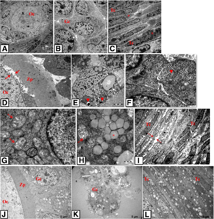
Transmission electron microscopy photomicrographs reveal ultrastructural details of oocytes, granulosa cells, and theca cells in control and PCOS groups. PCOS follicles exhibit mitochondrial swelling, dilated endoplasmic reticulum, and other signs of cellular stress.
Effect of resveratrol and metformin on ovarian reserve and ultrastructure in PCOS: …
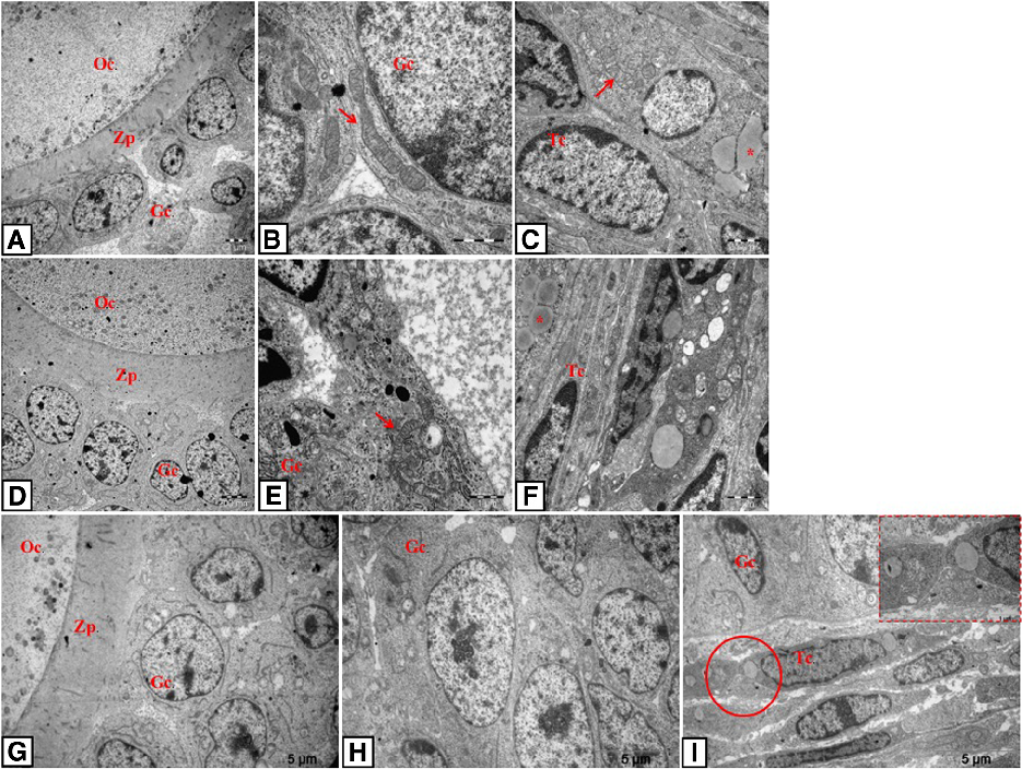
TEM photomicrographs of resveratrol-, metformin-, and combination-treated rat ovarian tissue show improved ultrastructural features compared to untreated PCOS. Mitochondrial morphology and endoplasmic reticulum integrity are partially restored.
Effect of resveratrol and metformin on ovarian reserve and ultrastructure in PCOS: …
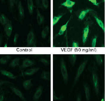
COX-2 protein expression is upregulated by VEGF stimulation in intestinal endothelial cells, and curcumin effectively blocks this induction. Prostaglandin E2 production follows a similar pattern of inhibition.
Curcumin inhibits VEGF-mediated angiogenesis in human intestinal microvascular endothelial cells through COX-2 …
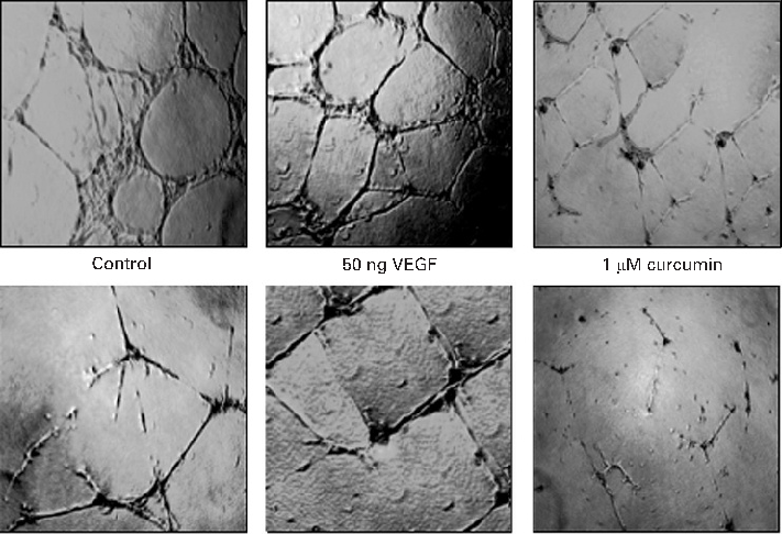
MAPK signaling pathway activation by VEGF is attenuated by curcumin in a time- and dose-dependent manner. Phosphorylation of ERK, p38, and JNK is markedly reduced in curcumin-treated endothelial cells.
Curcumin inhibits VEGF-mediated angiogenesis in human intestinal microvascular endothelial cells through COX-2 …
![Figure 1.Figure 1.Transmembrane orientation of astaxanthin [Transmembrane orientation of astaxanthin [1,9]. 1,9].](https://pdfs.citedhealth.com/figures/26861359/55.png)
Astaxanthin's transmembrane orientation is unique among carotenoids, spanning the entire lipid bilayer with its polar end groups anchored at both membrane surfaces. This positioning enables superior antioxidant protection of membrane lipids.
Potential Anti-Atherosclerotic Properties of Astaxanthin.
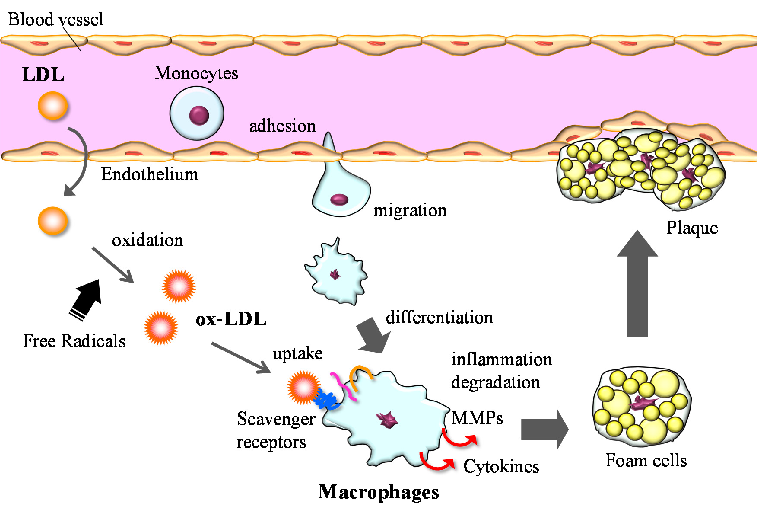
Macrophages play a central role in atherosclerosis development through uptake of oxidized LDL, foam cell formation, and secretion of inflammatory mediators. Astaxanthin may interrupt this process by reducing LDL oxidation and modulating macrophage activity.
Potential Anti-Atherosclerotic Properties of Astaxanthin.
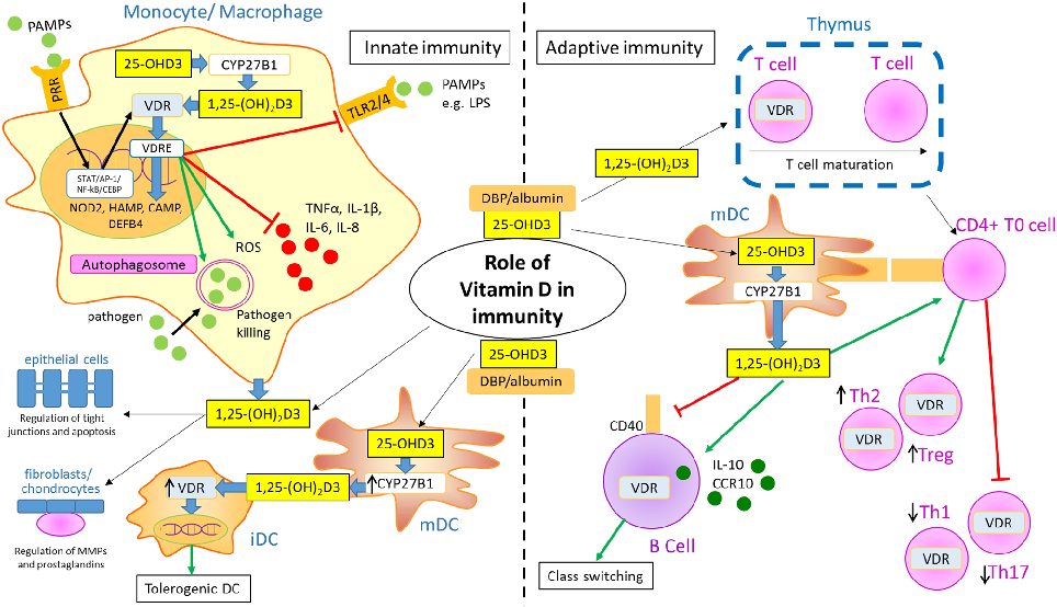
Vitamin D's immunomodulatory effects extend to multiple immune cell types relevant to rheumatoid arthritis, including T cells, B cells, dendritic cells, and macrophages. Vitamin D receptor activation promotes anti-inflammatory and tolerogenic immune phenotypes.
Vitamin D, Autoimmune Disease and Rheumatoid Arthritis.
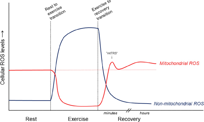
Proposed relative contributions of mitochondrial and non-mitochondrial ROS sources to overall cellular ROS levels in skeletal muscle during and after exercise are depicted. NADPH oxidase, xanthine oxidase, and mitochondrial electron transport chain are the primary generators.
Antioxidant supplements and endurance exercise: Current evidence and mechanistic insights.
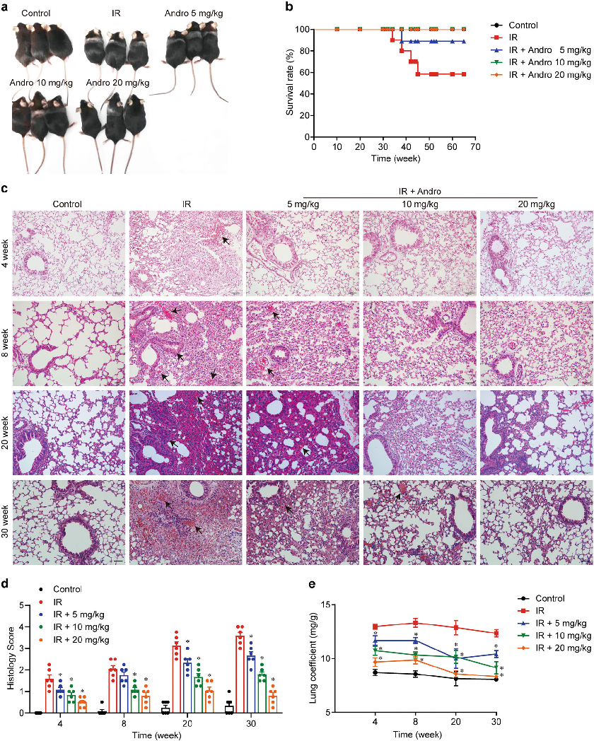
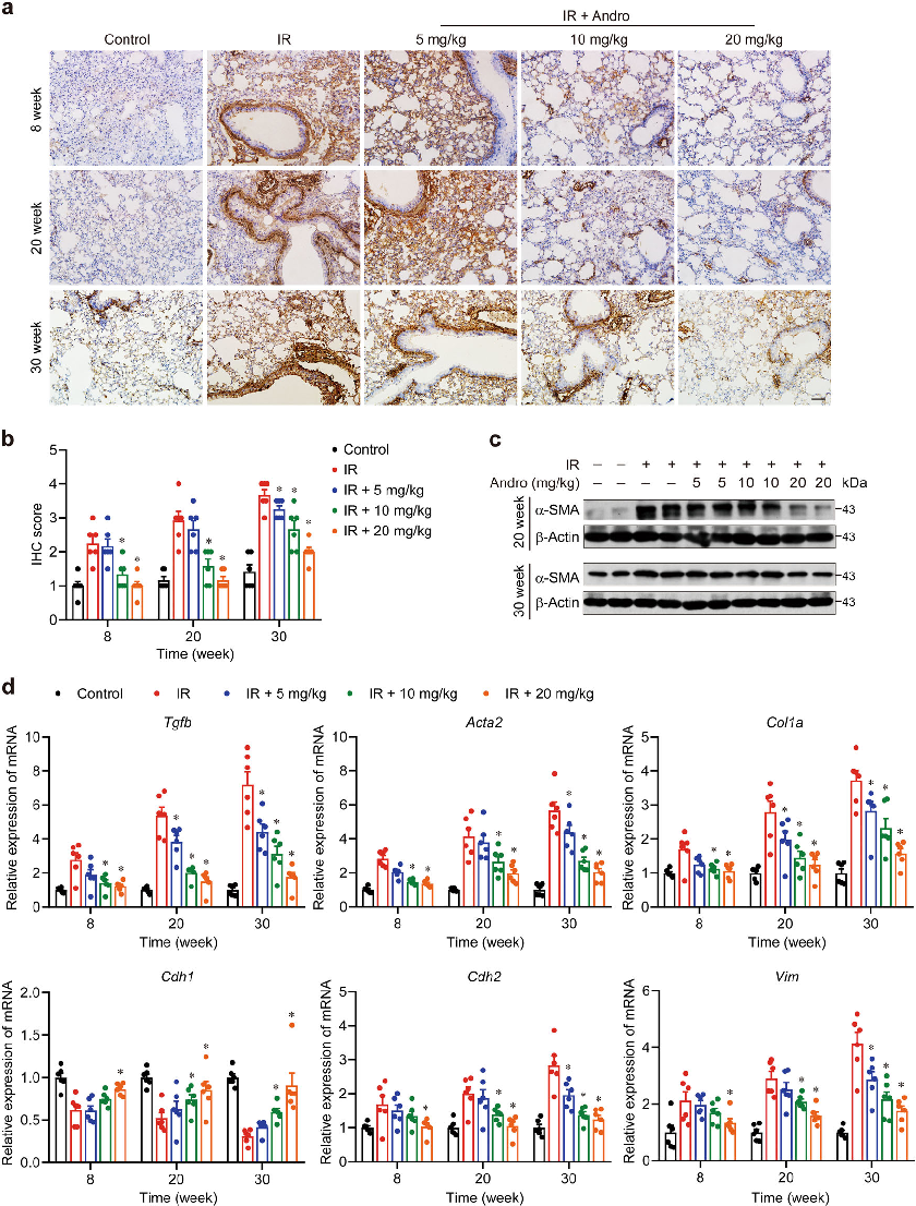
Mice exposed to 18 Gy irradiation and treated with varying doses of andrographolide for 4 weeks showed dose-dependent protection from radiation-induced lung injury. Representative images and quantitative data indicate that andrographolide significantly attenuated lung tissue damage.
Inhibition of AIM2 inflammasome-mediated pyroptosis by Andrographolide contributes to amelioration of radiation-induced …
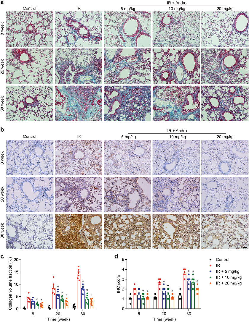
Histological analysis of lung tissue from irradiated mice reveals the extent of inflammatory cell infiltration and fibrosis. Andrographolide treatment appears to reduce these pathological changes in a dose-dependent manner.
Inhibition of AIM2 inflammasome-mediated pyroptosis by Andrographolide contributes to amelioration of radiation-induced …
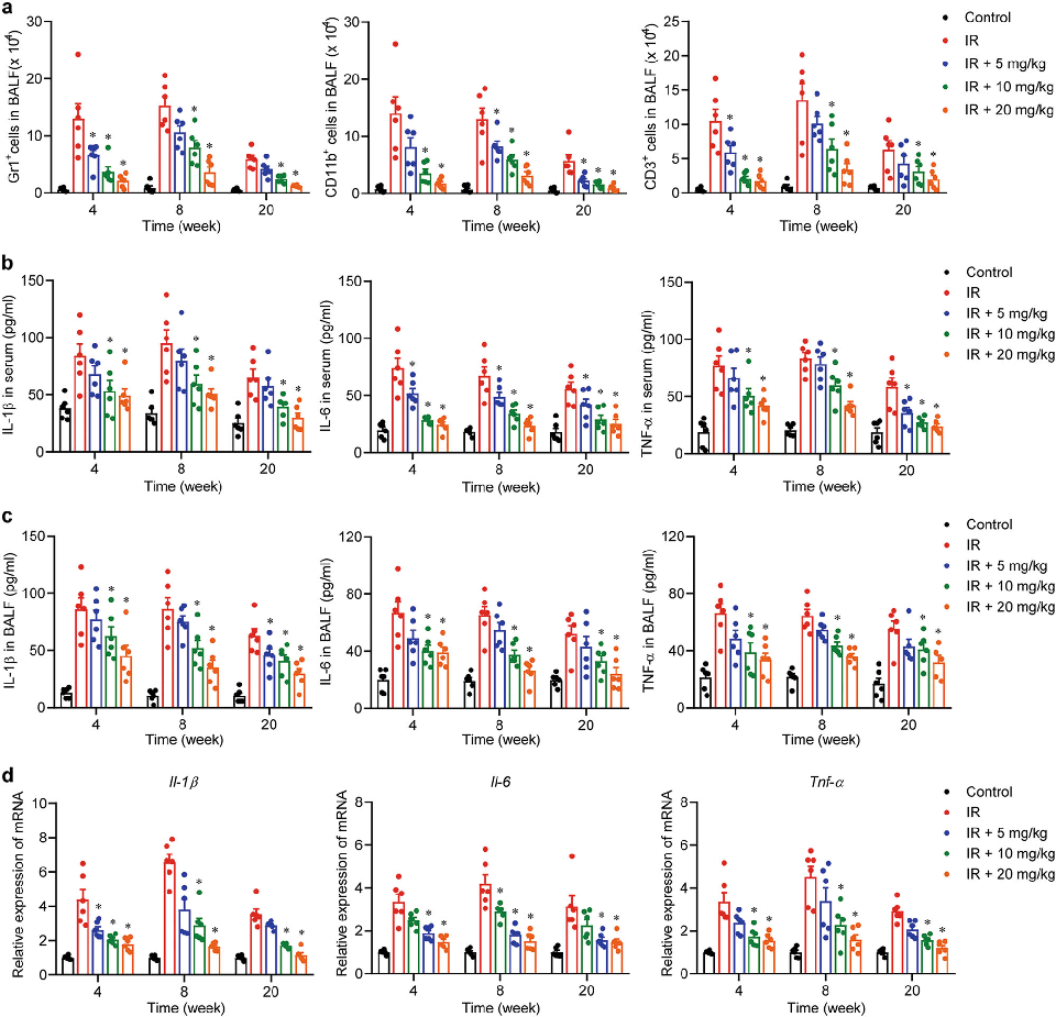
Inflammatory cytokine levels in lung tissue were measured following radiation exposure and andrographolide treatment. The data suggest that andrographolide suppresses pro-inflammatory mediator release in irradiated lung tissue.
Inhibition of AIM2 inflammasome-mediated pyroptosis by Andrographolide contributes to amelioration of radiation-induced …
AIM2 inflammasome activation and caspase-1-mediated pyroptosis play key roles in radiation-induced lung inflammation. This figure presents protein expression data showing andrographolide's inhibitory effects on the AIM2 inflammasome pathway.
Inhibition of AIM2 inflammasome-mediated pyroptosis by Andrographolide contributes to amelioration of radiation-induced …
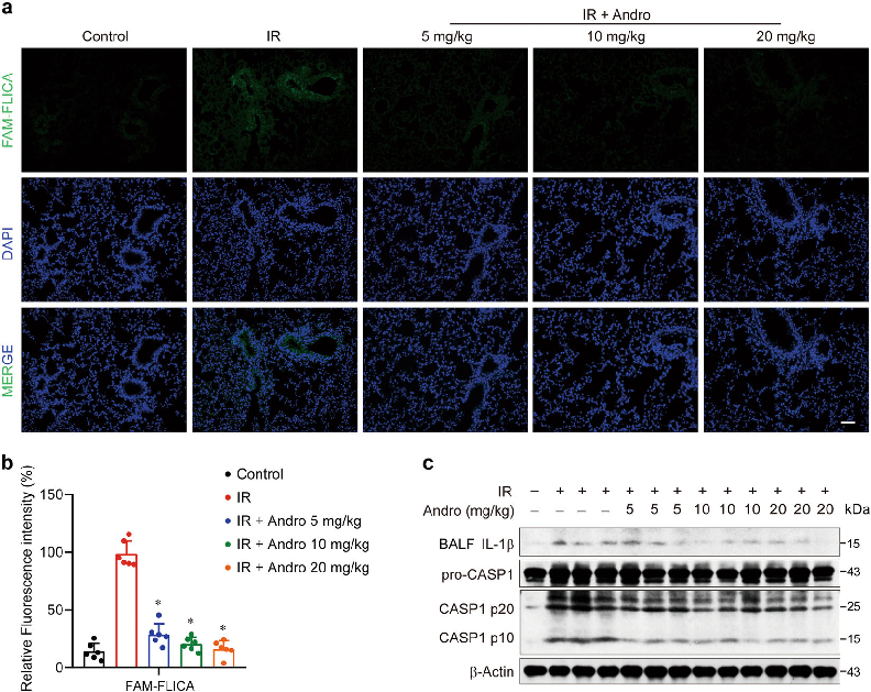
Gasdermin D cleavage is a downstream event in pyroptotic cell death triggered by radiation. Western blot analysis demonstrates that andrographolide reduces Gasdermin D processing in macrophages exposed to radiation.
Inhibition of AIM2 inflammasome-mediated pyroptosis by Andrographolide contributes to amelioration of radiation-induced …

Immunofluorescence or protein analysis reveals that andrographolide prevents AIM2 from binding cytoplasmic DNA and forming active inflammasome complexes. These findings indicate a specific molecular target for andrographolide's protective mechanism.
Inhibition of AIM2 inflammasome-mediated pyroptosis by Andrographolide contributes to amelioration of radiation-induced …
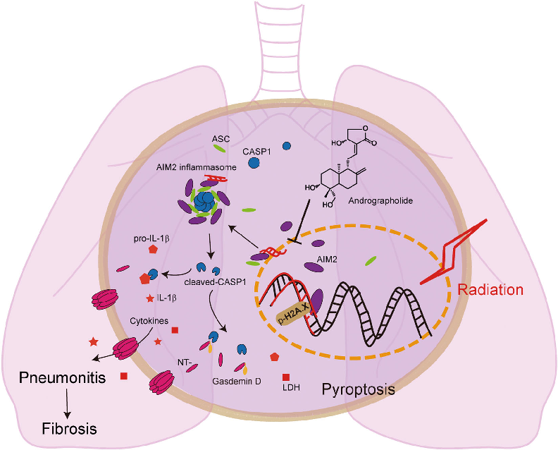
Andrographolide ameliorates radiation-induced lung injury by inhibiting Caspase-1-mediated Gasdermin D-dependent pyroptosis in macrophages. This schematic illustrates how the compound prevents AIM2 from translocating into the nucleus, thereby blocking inflammasome assembly and downstream inflammatory cascades.
Inhibition of AIM2 inflammasome-mediated pyroptosis by Andrographolide contributes to amelioration of radiation-induced …
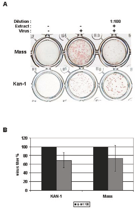
Standardized elderberry (Sambucus nigra) extract was tested against influenza virus propagation using a focus size reduction assay. MDCK cells infected with two influenza strains and incubated with the extract (1:100 dilution) showed reduced viral foci, indicating dose-dependent antiviral activity.
Inhibitory activity of a standardized elderberry liquid extract against clinically-relevant human respiratory …
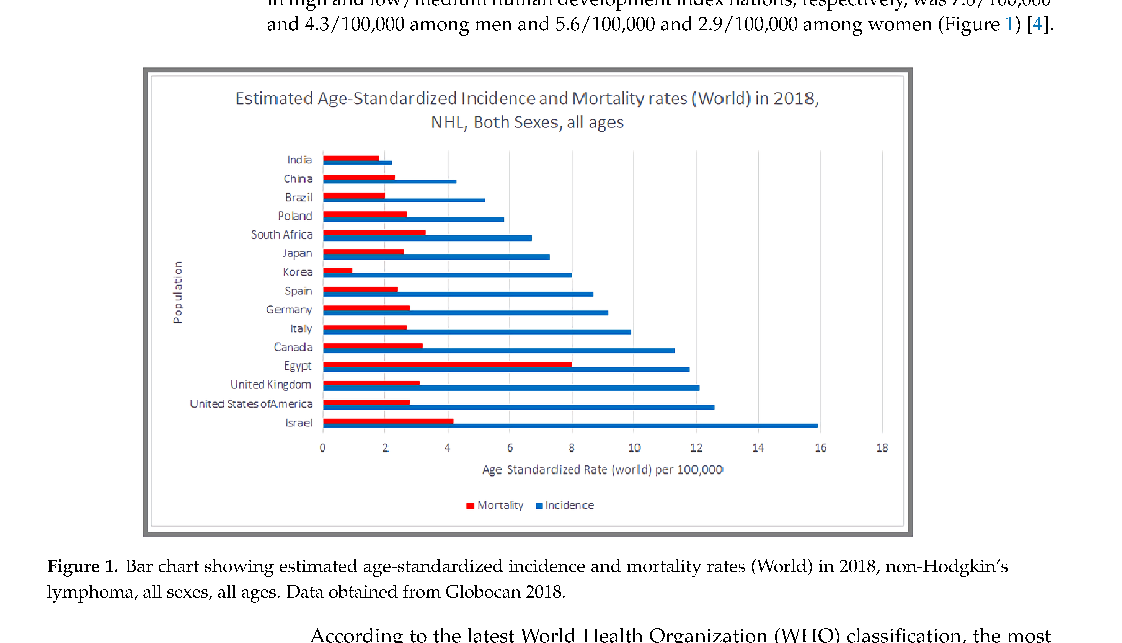
NHL mortality patterns reflect disparities in treatment access and disease subtype distribution. This figure presents survival and mortality data across different NHL classifications.
Epidemiology of Non-Hodgkin's Lymphoma.
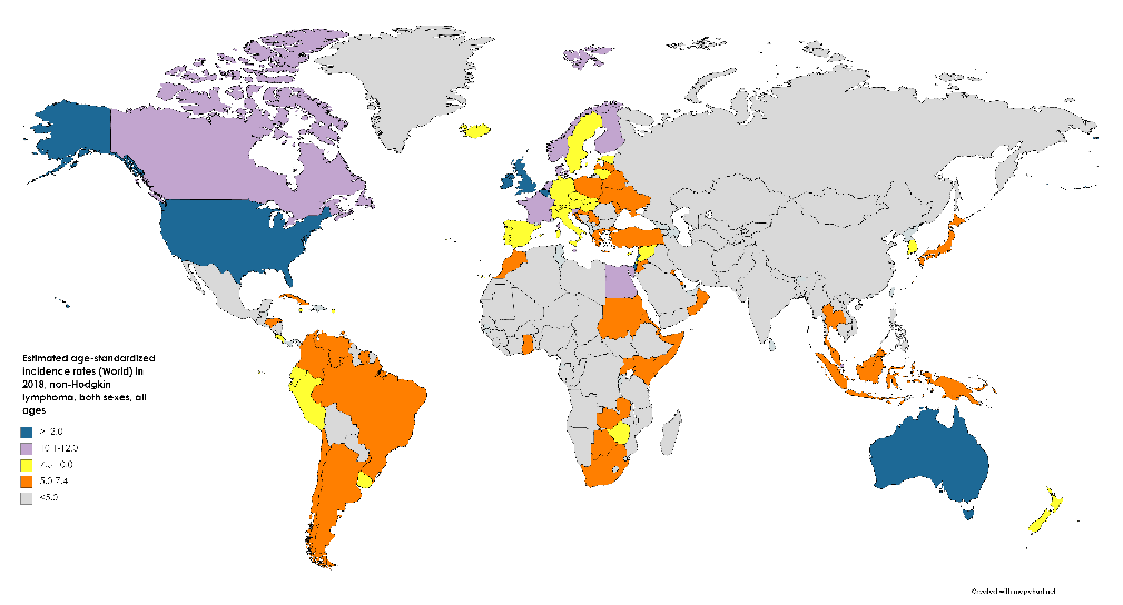
The epidemiological profile of Non-Hodgkin's lymphoma continues to evolve with improved molecular classification. This figure provides additional epidemiological data on NHL subtypes and their relative frequencies.
Epidemiology of Non-Hodgkin's Lymphoma.
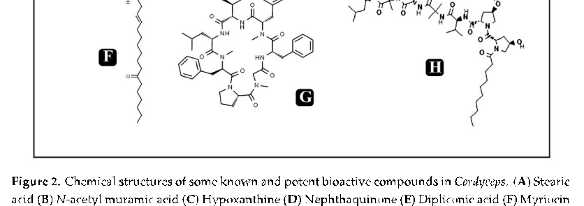
Neuroprotective and anti-fatigue properties of cordycepin have been demonstrated in preclinical models. This figure highlights cordycepin's potential benefits for neurological health and physical performance.
Cordycepin for Health and Wellbeing: A Potent Bioactive Metabolite of an Entomopathogenic …
Cordycepin modulates immune responses through effects on macrophage activation and cytokine production. This figure presents immunomodulatory data from in vitro and in vivo studies of cordycepin treatment.
Cordycepin for Health and Wellbeing: A Potent Bioactive Metabolite of an Entomopathogenic …
Anti-microbial and anti-viral activities of cordycepin complement its anti-inflammatory properties. This figure summarizes evidence for cordycepin's broad-spectrum antimicrobial potential.
Cordycepin for Health and Wellbeing: A Potent Bioactive Metabolite of an Entomopathogenic …
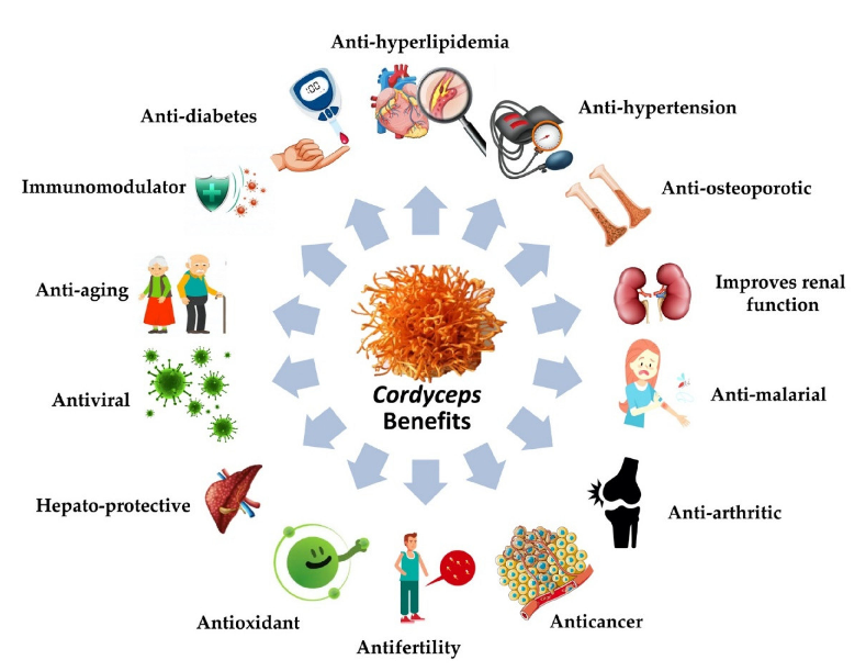
Cordyceps-derived products are available globally as nutraceutical supplements. This figure combines a pictorial representation of Cordyceps' therapeutic potential with a table of commercially available nutraceutical formulations.
Cordycepin for Health and Wellbeing: A Potent Bioactive Metabolite of an Entomopathogenic …
第 2 页,共 14 页