研究流程
117 来自同行评审研究的图表
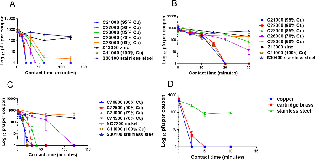
Viral titer decay curves on stainless steel and other metal surfaces demonstrate that coronavirus 229E can remain infectious for several days under ambient conditions.
Human Coronavirus 229E Remains Infectious on Common Touch Surface Materials.
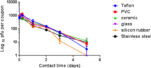
Comparison of coronavirus inactivation rates across polymer-based surfaces including PVC, silicone rubber, and Teflon shows material-dependent variation in viral survival.
Human Coronavirus 229E Remains Infectious on Common Touch Surface Materials.
Temperature and humidity effects on coronavirus 229E survival on surfaces are displayed, with higher temperatures generally associated with faster viral inactivation.
Human Coronavirus 229E Remains Infectious on Common Touch Surface Materials.
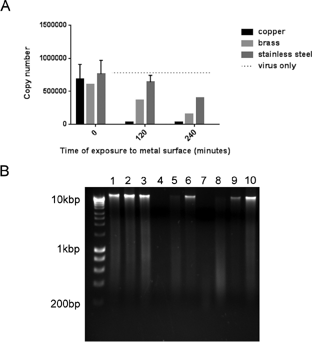
Recovery efficiency controls for the surface sampling methodology are shown, validating the quantitative viral titer measurements across different material types.
Human Coronavirus 229E Remains Infectious on Common Touch Surface Materials.
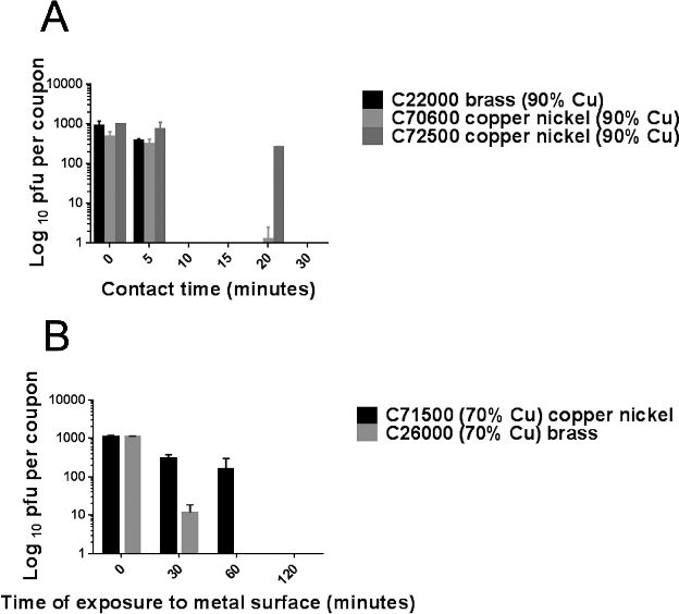
Summary comparison of coronavirus 229E persistence across all tested surface materials, ranked by duration of detectable infectivity.
Human Coronavirus 229E Remains Infectious on Common Touch Surface Materials.
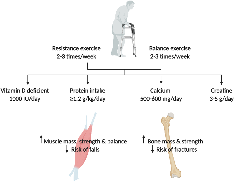
Outcome data from intervention studies targeting both bone and muscle health in osteosarcopenic patients are compared across different therapeutic approaches.
Osteosarcopenia: epidemiology, diagnosis, and treatment-facts and numbers.
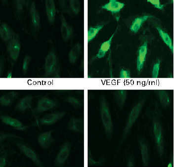
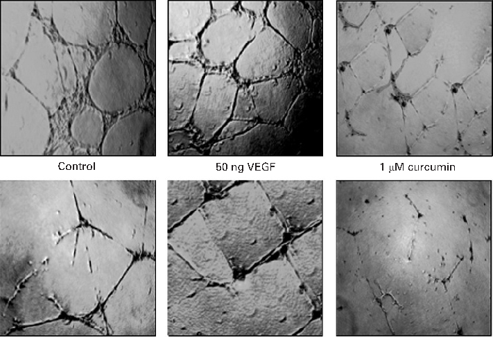
COX-2 protein expression is upregulated by VEGF stimulation in intestinal endothelial cells, and curcumin effectively blocks this induction. Prostaglandin E2 production follows a similar pattern of inhibition.
Curcumin inhibits VEGF-mediated angiogenesis in human intestinal microvascular endothelial cells through COX-2 …
MAPK signaling pathway activation by VEGF is attenuated by curcumin in a time- and dose-dependent manner. Phosphorylation of ERK, p38, and JNK is markedly reduced in curcumin-treated endothelial cells.
Curcumin inhibits VEGF-mediated angiogenesis in human intestinal microvascular endothelial cells through COX-2 …
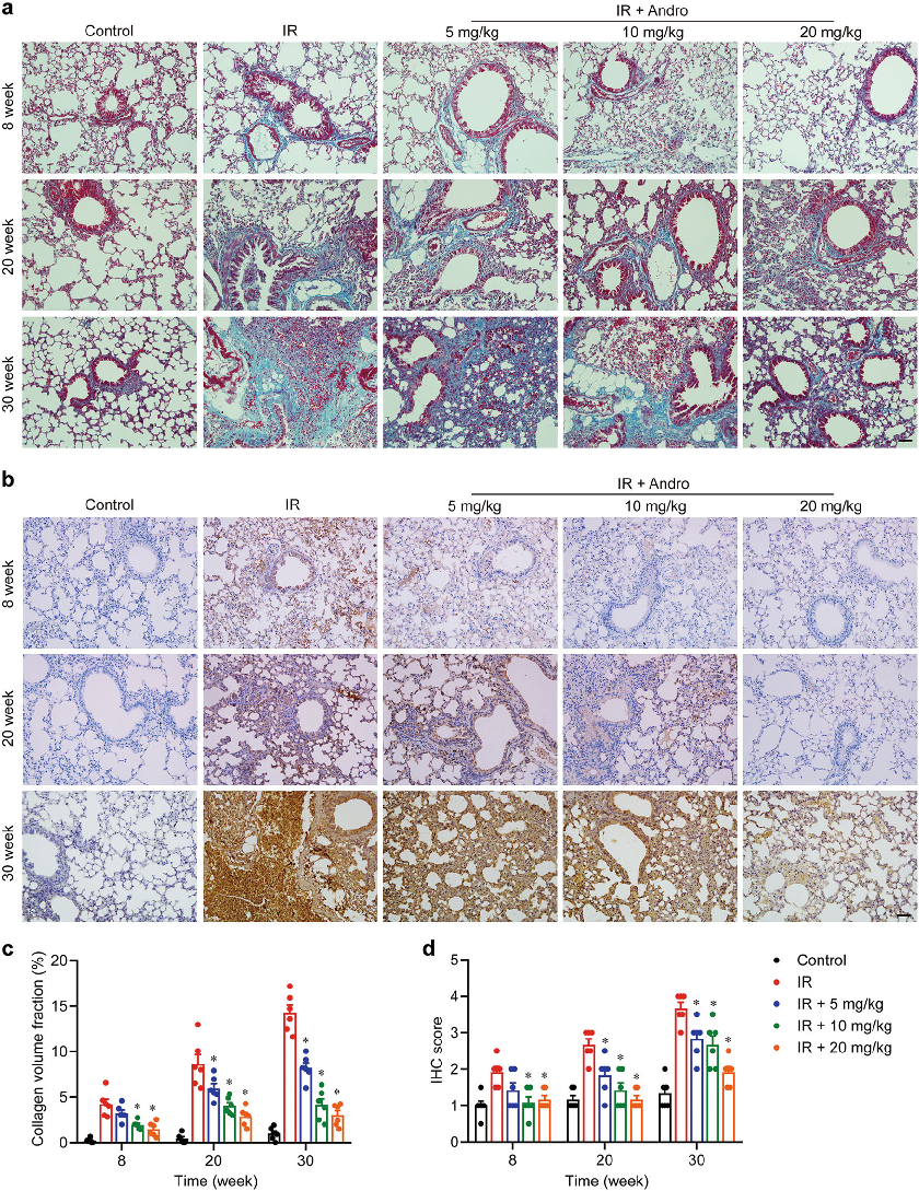
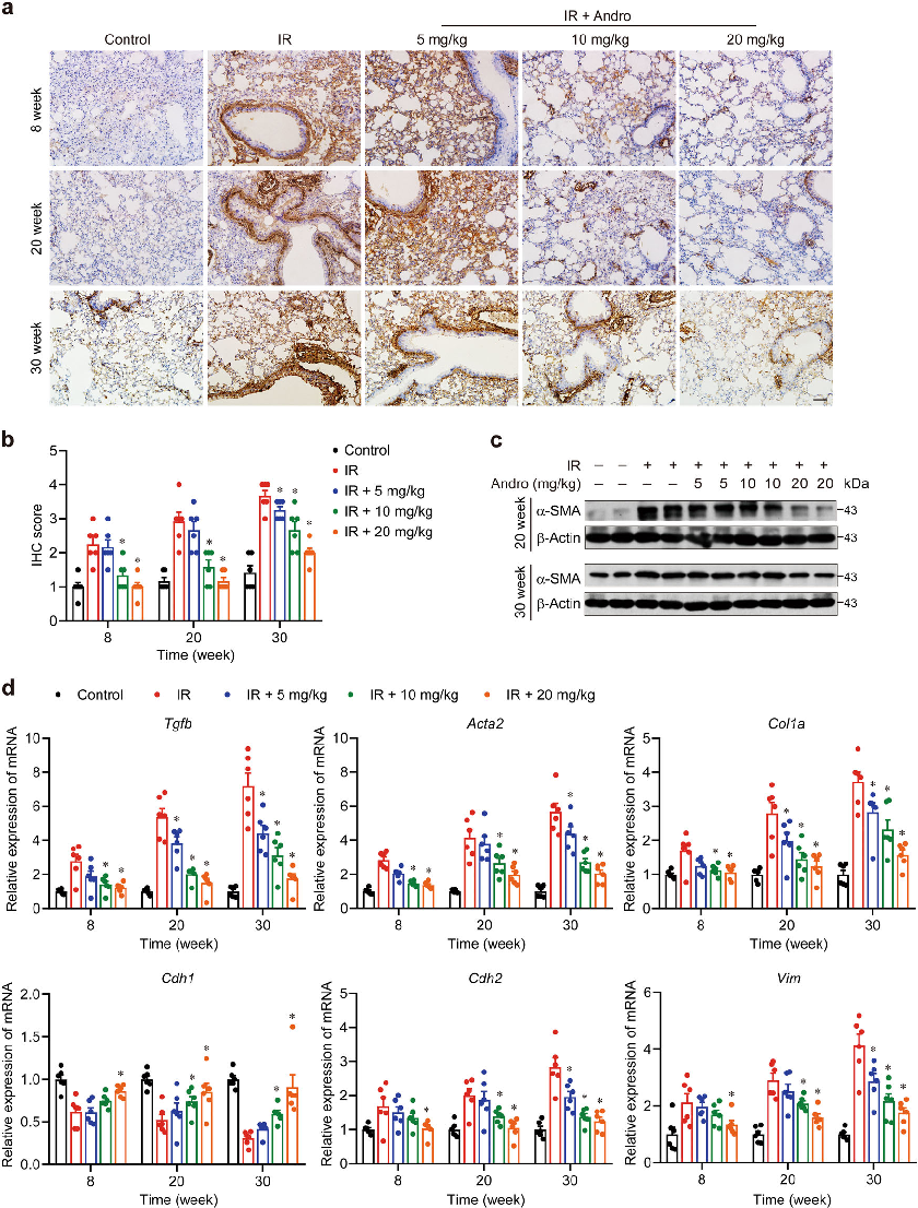
Inflammatory cytokine levels in lung tissue were measured following radiation exposure and andrographolide treatment. The data suggest that andrographolide suppresses pro-inflammatory mediator release in irradiated lung tissue.
Inhibition of AIM2 inflammasome-mediated pyroptosis by Andrographolide contributes to amelioration of radiation-induced …
AIM2 inflammasome activation and caspase-1-mediated pyroptosis play key roles in radiation-induced lung inflammation. This figure presents protein expression data showing andrographolide's inhibitory effects on the AIM2 inflammasome pathway.
Inhibition of AIM2 inflammasome-mediated pyroptosis by Andrographolide contributes to amelioration of radiation-induced …
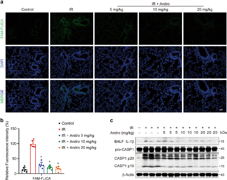
Gasdermin D cleavage is a downstream event in pyroptotic cell death triggered by radiation. Western blot analysis demonstrates that andrographolide reduces Gasdermin D processing in macrophages exposed to radiation.
Inhibition of AIM2 inflammasome-mediated pyroptosis by Andrographolide contributes to amelioration of radiation-induced …

Immunofluorescence or protein analysis reveals that andrographolide prevents AIM2 from binding cytoplasmic DNA and forming active inflammasome complexes. These findings indicate a specific molecular target for andrographolide's protective mechanism.
Inhibition of AIM2 inflammasome-mediated pyroptosis by Andrographolide contributes to amelioration of radiation-induced …
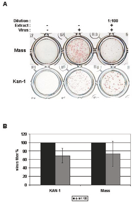
Standardized elderberry (Sambucus nigra) extract was tested against influenza virus propagation using a focus size reduction assay. MDCK cells infected with two influenza strains and incubated with the extract (1:100 dilution) showed reduced viral foci, indicating dose-dependent antiviral activity.
Inhibitory activity of a standardized elderberry liquid extract against clinically-relevant human respiratory …
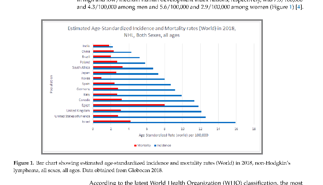
NHL mortality patterns reflect disparities in treatment access and disease subtype distribution. This figure presents survival and mortality data across different NHL classifications.
Epidemiology of Non-Hodgkin's Lymphoma.
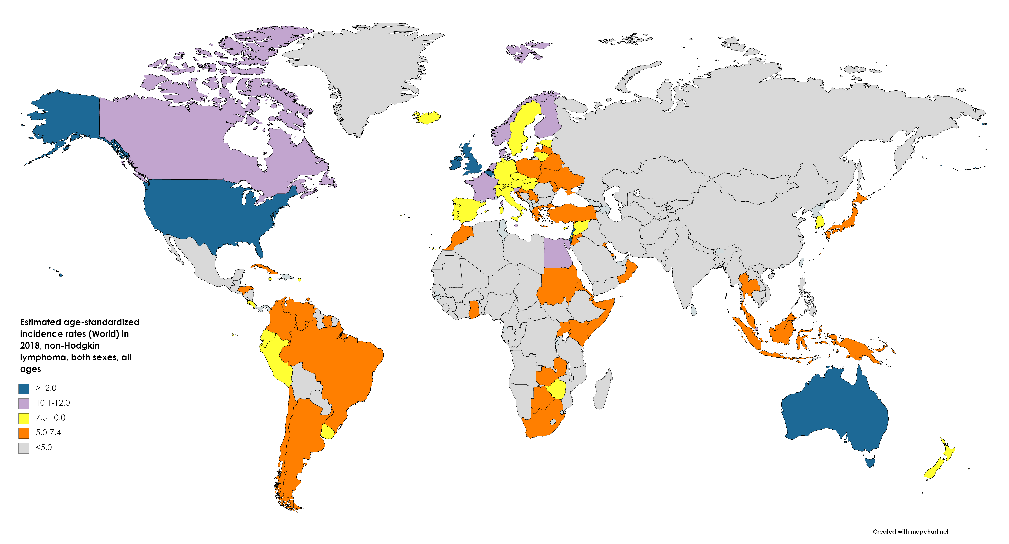
The epidemiological profile of Non-Hodgkin's lymphoma continues to evolve with improved molecular classification. This figure provides additional epidemiological data on NHL subtypes and their relative frequencies.
Epidemiology of Non-Hodgkin's Lymphoma.
![Figure 5. Expected global nutraceutical market by 2025 with China, India, Tibet and Nepal as global leaders for production and extraction of cordycepin [144].](https://pdfs.citedhealth.com/figures/32545666/223.png)
Global demand for Cordyceps-based nutraceuticals is concentrated in China, India, Tibet, and Nepal. This figure projects the expected nutraceutical market growth, with these regions positioned as global leaders in cordycepin production and extraction.
Cordycepin for Health and Wellbeing: A Potent Bioactive Metabolite of an Entomopathogenic …
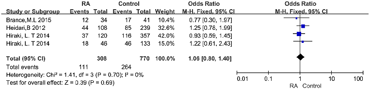
Geographic and seasonal variation in vitamin D status may confound the relationship with RA disease activity. This figure presents stratified analyses accounting for latitude, season, or supplementation status among RA patients.
Serum Vitamin D Level and Rheumatoid Arthritis Disease Activity: Review and Meta-Analysis.
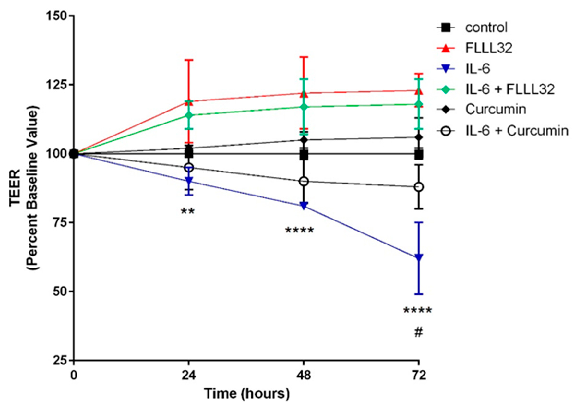
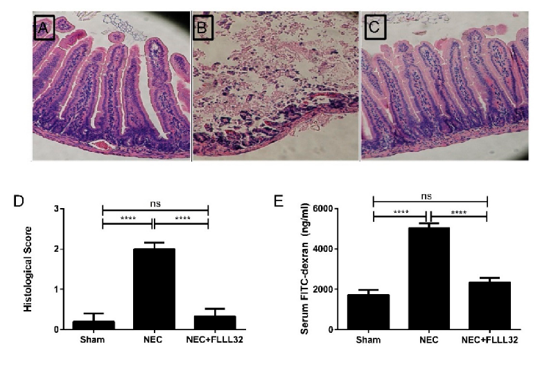
FLLL32 (a curcumin analogue) and curcumin both protect against IL-6-induced reduction of transepithelial electrical resistance (TEER) in T84 cell monolayers. This graph shows that curcumin preserves intestinal barrier integrity by counteracting cytokine-mediated tight junction disruption over 72 hours.
Curcumin and Intestinal Inflammatory Diseases: Molecular Mechanisms of Protection.
Curcumin's effects on intestinal tight junction proteins have been demonstrated in multiple experimental models. This figure presents protein expression or immunofluorescence data showing curcumin-mediated preservation of barrier function.
Curcumin and Intestinal Inflammatory Diseases: Molecular Mechanisms of Protection.
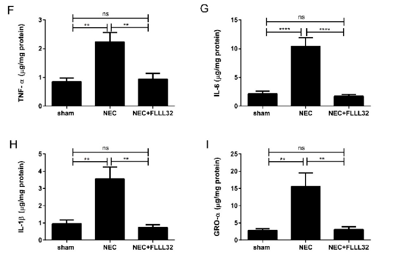
Animal models of necrotizing enterocolitis and colitis respond favorably to curcumin treatment. This figure presents in vivo data on curcumin's protective effects in experimental intestinal inflammation.
Curcumin and Intestinal Inflammatory Diseases: Molecular Mechanisms of Protection.
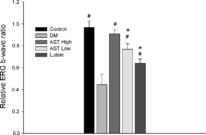
Electroretinography (ERG) recordings evaluating retinal function in control and STZ-induced diabetic rats treated with normal saline, 0.6 mg/kg AST, 3 mg/kg AST, or 0.5 mg/kg lutein for 8 weeks. ERG wave amplitudes indicate the functional impact of each treatment on diabetic retinal physiology.
Astaxanthin Inhibits Expression of Retinal Oxidative Stress and Inflammatory Mediators in Streptozotocin-Induced …
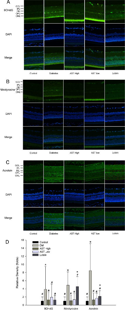
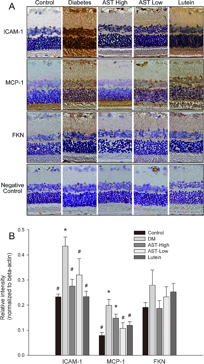
Inflammatory marker expression levels in retinal tissue or aqueous humor of diabetic rats, comparing astaxanthin-treated groups with untreated diabetic controls. Results indicate AST may modulate inflammatory mediator production.
Astaxanthin Inhibits Expression of Retinal Oxidative Stress and Inflammatory Mediators in Streptozotocin-Induced …
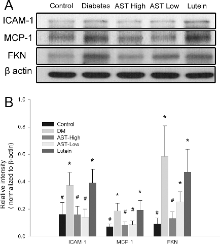
Western blot or protein expression analysis of retinal inflammatory and oxidative stress markers in the diabetic rat model, demonstrating the molecular effects of astaxanthin treatment on key signaling proteins.
Astaxanthin Inhibits Expression of Retinal Oxidative Stress and Inflammatory Mediators in Streptozotocin-Induced …
Gene or protein expression data for retinal vascular endothelial growth factor (VEGF) or related angiogenic markers in AST-treated versus untreated diabetic rats.
Astaxanthin Inhibits Expression of Retinal Oxidative Stress and Inflammatory Mediators in Streptozotocin-Induced …
第 1 页,共 5 页