अनुसंधान प्रक्रिया
333 सहकर्मी-समीक्षित शोध से आंकड़े
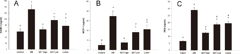
Quantification of ICAM-1, MCP-1, and fractalkine (FKN) protein levels in aqueous humor of diabetic rats treated with astaxanthin or lutein. Reduced expression of these inflammatory mediators suggests AST may attenuate vascular inflammation in diabetic eyes.
Astaxanthin Inhibits Expression of Retinal Oxidative Stress and Inflammatory Mediators in Streptozotocin-Induced …
Supplementary analysis of inflammatory or oxidative markers in ocular tissues of STZ-induced diabetic rats, providing additional evidence for astaxanthin's protective mechanism against diabetic retinal damage.
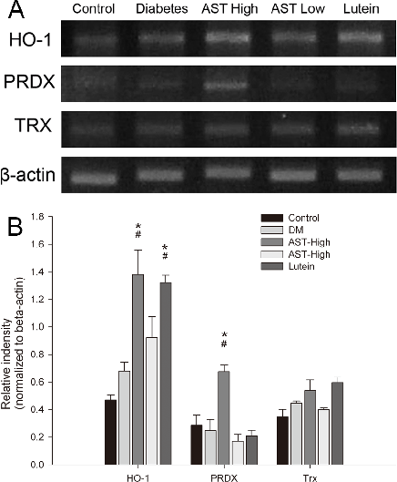
Astaxanthin Inhibits Expression of Retinal Oxidative Stress and Inflammatory Mediators in Streptozotocin-Induced …
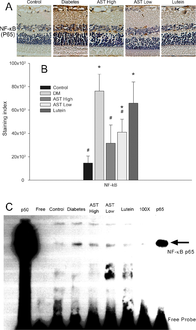
Summary of retinal protection outcomes or dose-response data for astaxanthin in the diabetic rat model, consolidating evidence that AST inhibits expression of oxidative stress and inflammatory mediators in diabetic retinopathy.
Astaxanthin Inhibits Expression of Retinal Oxidative Stress and Inflammatory Mediators in Streptozotocin-Induced …
Tabular or graphical summary of animal model studies evaluating resveratrol's therapeutic effects on endometriotic lesion size, vascularization, and inflammatory marker expression.
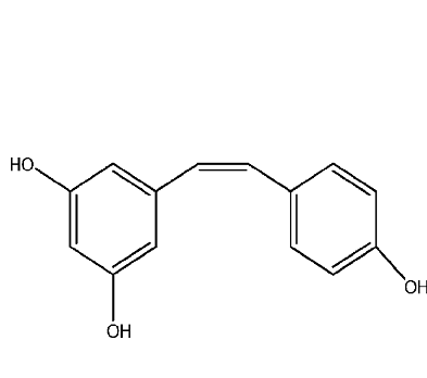
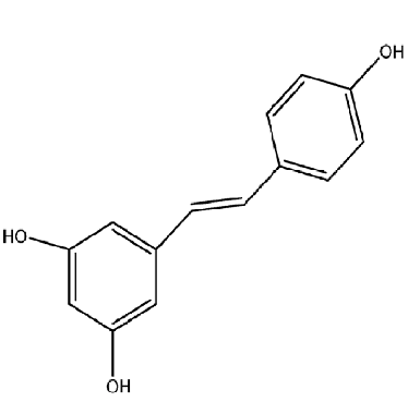
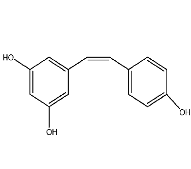
Therapeutic Approaches of Resveratrol on Endometriosis via Anti-Inflammatory and Anti-Angiogenic Pathways.
Molecular mechanisms underlying resveratrol's anti-inflammatory activity in endometriosis, including NF-kB inhibition and cytokine modulation pathways.
Therapeutic Approaches of Resveratrol on Endometriosis via Anti-Inflammatory and Anti-Angiogenic Pathways.
Anti-angiogenic effects of resveratrol in endometriosis models, showing inhibition of VEGF expression or microvessel density in endometriotic implants.
Therapeutic Approaches of Resveratrol on Endometriosis via Anti-Inflammatory and Anti-Angiogenic Pathways.
Comparison of resveratrol with conventional endometriosis therapies or other natural compounds, highlighting potential advantages of the polyphenolic approach.
Therapeutic Approaches of Resveratrol on Endometriosis via Anti-Inflammatory and Anti-Angiogenic Pathways.
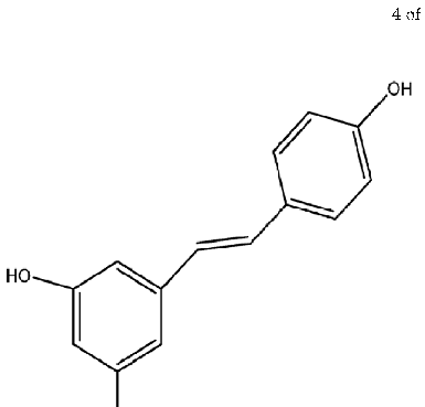
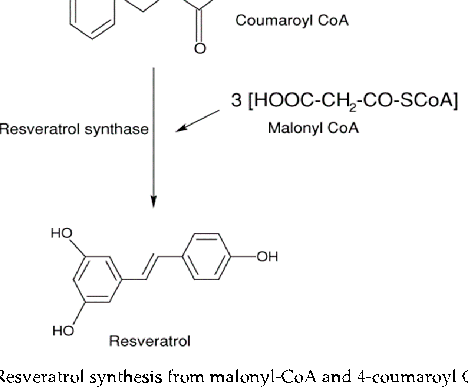
![Figure 2. Resveratrol synthesis from malonyl-CoA and 4-coumaroyl CoA [43].](https://pdfs.citedhealth.com/figures/30781885/114.png)
Biosynthetic pathway of resveratrol from malonyl-CoA and 4-coumaroyl CoA precursors. Understanding the natural synthesis route informs efforts to produce this polyphenol for therapeutic applications in conditions such as endometriosis.
Therapeutic Approaches of Resveratrol on Endometriosis via Anti-Inflammatory and Anti-Angiogenic Pathways.
Detailed analysis of resveratrol's molecular targets in endometrial tissue, focusing on oxidative stress reduction and apoptosis induction in ectopic endometrial cells.
Therapeutic Approaches of Resveratrol on Endometriosis via Anti-Inflammatory and Anti-Angiogenic Pathways.
![Figure 3. Illustration of molecular pathways in the development of endometriosis [7,68,73–75].](https://pdfs.citedhealth.com/figures/30781885/143.png)
Comprehensive pathway illustration showing the molecular mechanisms involved in endometriosis development, including estrogen signaling, inflammatory cascades, and angiogenic processes that resveratrol may therapeutically target.
Therapeutic Approaches of Resveratrol on Endometriosis via Anti-Inflammatory and Anti-Angiogenic Pathways.
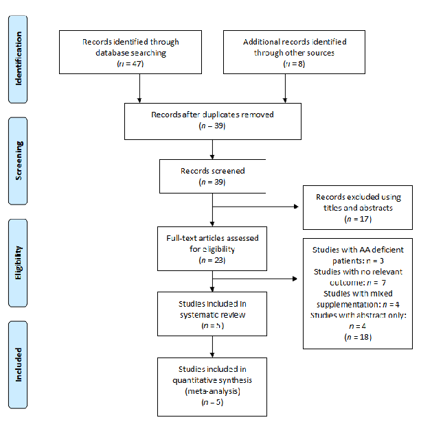
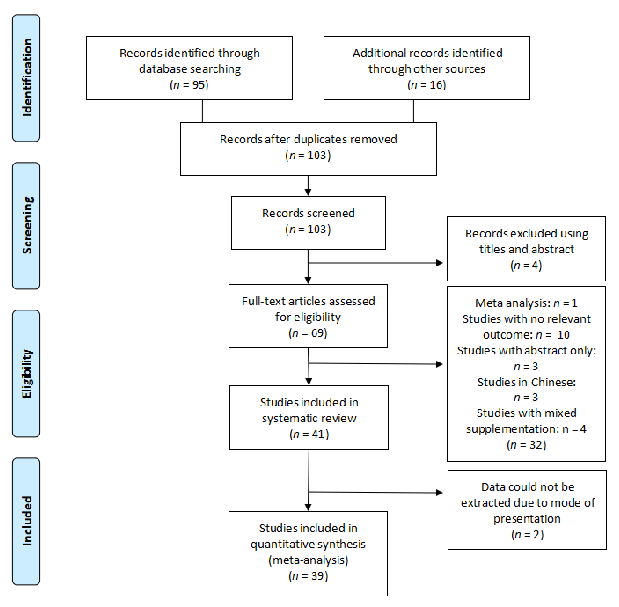
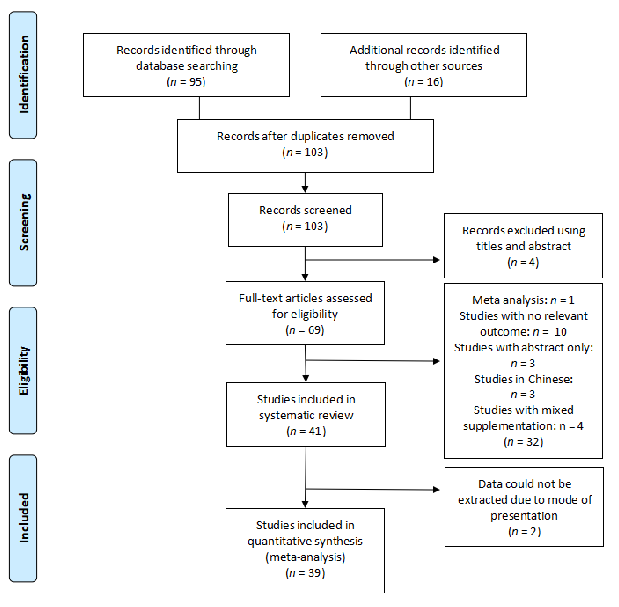
PRISMA flow diagram details the screening and selection process for studies on glutamine supplementation and wound healing. From the initial database search, studies were filtered through title screening, abstract review, and full-text assessment.
The Effect of Amino Acids on Wound Healing: A Systematic Review and …
Characteristics of included studies on arginine supplementation for wound healing are summarized in tabular form. The table captures study design, participant demographics, dosage protocols, and primary outcome measures across the selected trials.
The Effect of Amino Acids on Wound Healing: A Systematic Review and …
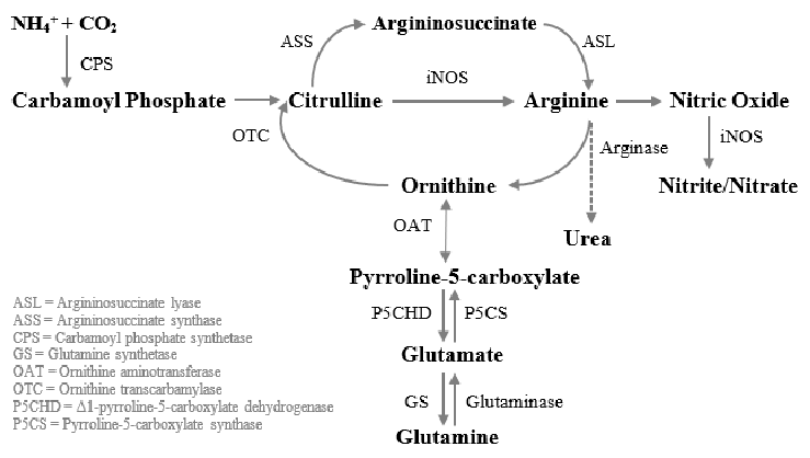
Glutamine-to-arginine metabolism in human macrophages is mapped, showing the conversion of carbamoyl phosphate and ornithine to citrulline via ornithine transcarbamylase, and subsequent transformation to argininosuccinate. This pathway is central to understanding how glutamine supports immune-mediated wound repair.
The Effect of Amino Acids on Wound Healing: A Systematic Review and …
Study characteristics for the glutamine supplementation arm of the systematic review are presented. The compilation enables cross-study comparison of dosing regimens, wound types, and healing outcomes measured in human trials.
The Effect of Amino Acids on Wound Healing: A Systematic Review and …
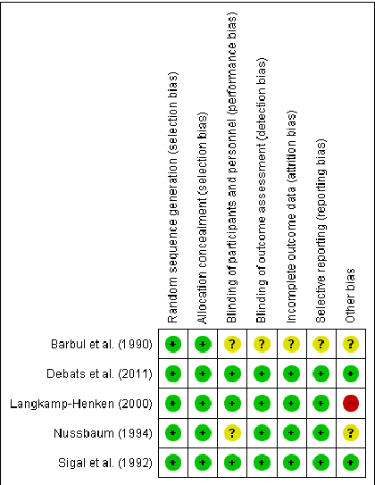
Risk of bias assessment for arginine studies is displayed using the Cochrane tool framework. Each domain of potential bias is evaluated across the included trials to gauge the overall quality of evidence supporting arginine supplementation for wound healing.
The Effect of Amino Acids on Wound Healing: A Systematic Review and …
Risk of bias assessment for glutamine studies complements the arginine evaluation. The systematic assessment of selection, performance, detection, attrition, and reporting bias helps contextualize the strength of conclusions about glutamine and wound repair.
The Effect of Amino Acids on Wound Healing: A Systematic Review and …
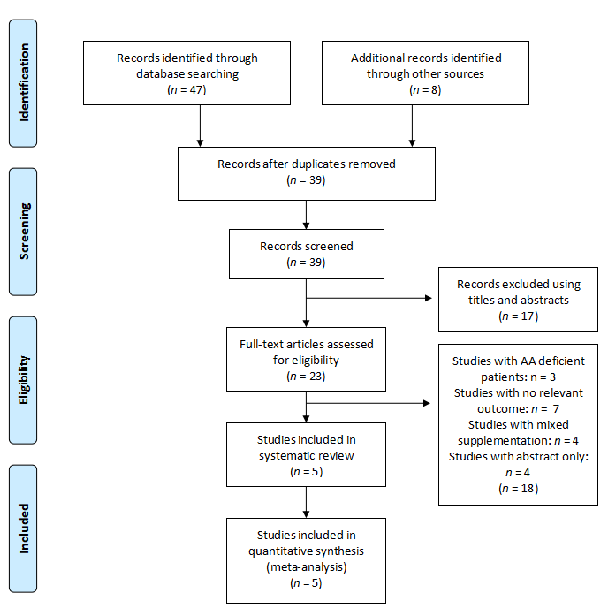
The PRISMA flow diagram for the arginine search strategy traces article identification through database queries, duplicate removal, and eligibility screening. Five human studies on arginine supplementation and wound healing met the final inclusion criteria.
The Effect of Amino Acids on Wound Healing: A Systematic Review and …
Risk of bias is summarized for each included arginine study using green (low risk), yellow (unclear), and red (high risk) indicators. The visual matrix helps readers quickly assess methodological quality across randomization, blinding, and outcome reporting domains.
The Effect of Amino Acids on Wound Healing: A Systematic Review and …
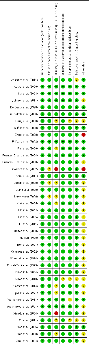
Risk of bias for the included glutamine studies follows the same Cochrane framework, with symbols indicating low, unclear, and high risk across multiple domains. The assessment covers 39 human studies evaluating glutamine supplementation effects on wound healing.
The Effect of Amino Acids on Wound Healing: A Systematic Review and …
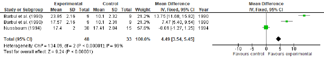
A fixed-effects meta-analysis forest plot displays the effect of arginine supplementation on hydroxyproline content, a biomarker of collagen synthesis in wound tissue. Individual study effect sizes and confidence intervals are pooled to estimate the overall treatment effect.
The Effect of Amino Acids on Wound Healing: A Systematic Review and …
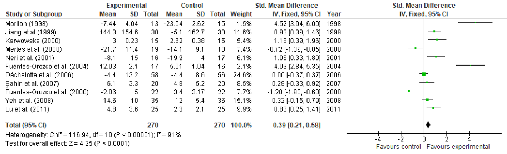
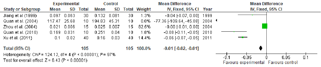
Nitrogen balance outcomes from glutamine supplementation studies are combined in a fixed-effects forest plot. Positive nitrogen balance indicates enhanced protein retention, which is associated with improved wound healing capacity in stressed or surgical patients.
The Effect of Amino Acids on Wound Healing: A Systematic Review and …
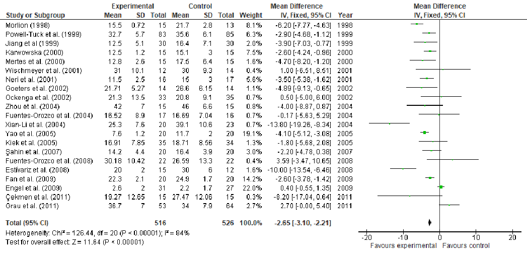
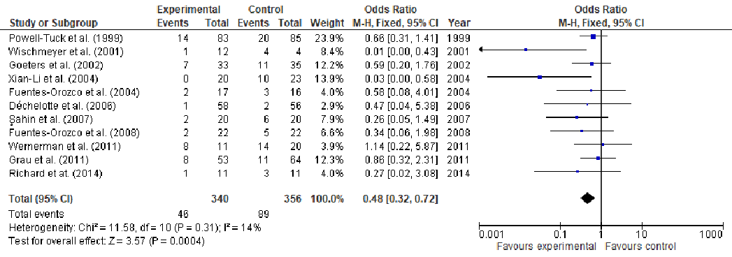
Length of hospital stay is analyzed in a fixed-effects meta-analysis forest plot for glutamine supplementation trials. The pooled estimate indicates whether glutamine may reduce recovery time in patients with wounds requiring hospitalization.
The Effect of Amino Acids on Wound Healing: A Systematic Review and …
Additional meta-analysis results for glutamine supplementation and wound healing outcomes are presented. The analysis extends the evidence base by examining secondary endpoints across the included clinical trials.
The Effect of Amino Acids on Wound Healing: A Systematic Review and …
Supplementary forest plot data from the glutamine meta-analysis displays effect estimates for an additional wound healing outcome. Confidence intervals for individual studies and the pooled effect are shown.
The Effect of Amino Acids on Wound Healing: A Systematic Review and …
पृष्ठ 4 / 14